
Calculate Volumes with VOI in the PET/CT Fusion Viewer
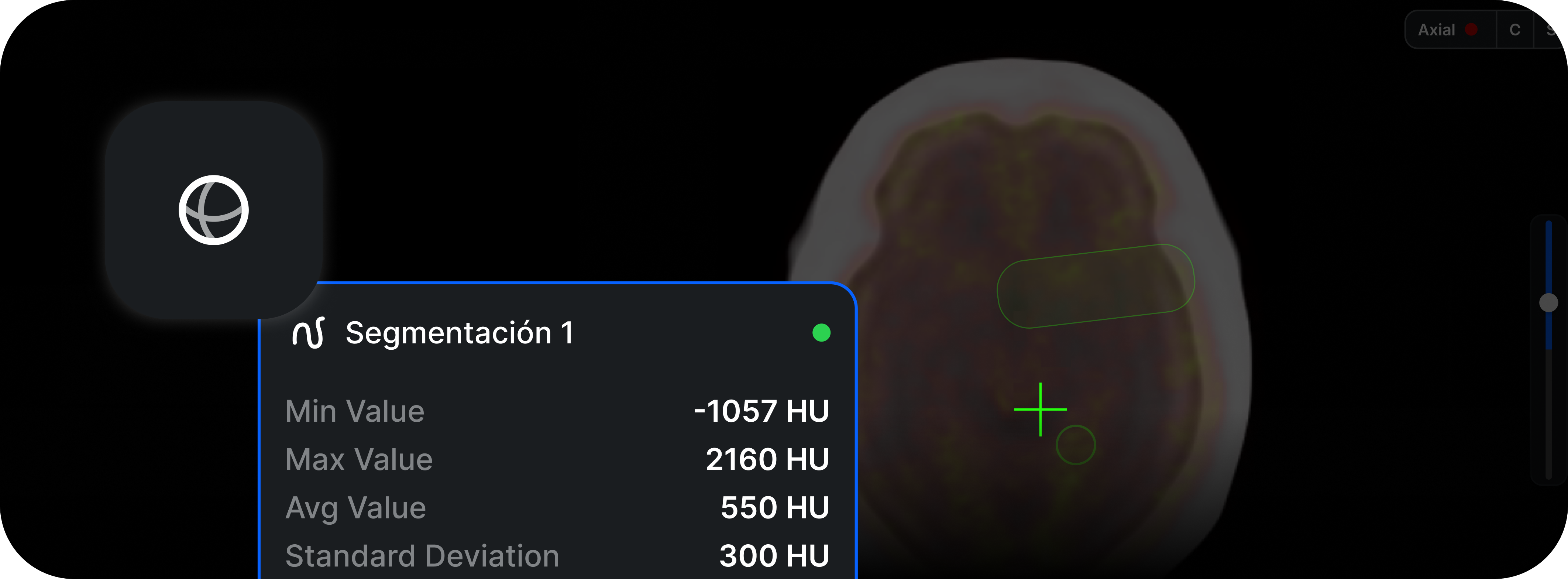
As part of strengthening the viewer's advanced capabilities for Nuclear Medicine, the ability to create, edit, and analyze VOIs (Volumes of Interest) directly on hybrid SPECT-CT and PET-CT studies has been added. This tool enables more precise quantitative measurements and integrated functional-anatomical characterization.
What it is
The VOI tool allows the user to delimit three-dimensional volumes on the functional images (SPECT or PET) and simultaneously view their anatomical correlation on CT. The generated VOIs can calculate basic statistics such as maximum, minimum, average, and approximate volume, depending on the data available in the study. The tool supports spherical, rectangular, and free-form (3D) VOIs, with visualization in multiple planes.
What it is for
The tool is useful for:
- Quantitative characterization of uptake foci.
- Lesion tracking in comparative studies.
- Consistent measurements in PET and SPECT-CT on equipment that exports quantitative data.
- Support for radiologists and nuclear medicine physicians in assessing therapeutic response.
- Improving the accuracy of integrated functional-anatomical readings.
How it affects the workflow
Previously, the user had to take 2D measurements or resort to external post-processing software. With this improvement, volumetric analysis is performed directly in the PACS viewer, aligned with the CT and without additional steps. VOIs can be moved, adjusted, or deleted, and remain visible while the user works within the same study. They are also integrated into the existing measurement workflow without modifying previous tools.
What changes compared to previous versions
Prior to this update, the viewer only allowed point or linear measurements on Nuclear Medicine studies. There was no native VOI capability for SPECT-CT, and the tool was limited to PET-CT in basic modes.
Now:
- VOI is enabled for both PET-CT and SPECT-CT.
- Integration with CT for anatomical reference is improved.
- Basic volumetric statistics are added where the equipment data allow it.
- Performance when manipulating 3D volumes is optimized.
Implications for the user
VOI availability depends on the quality of the study and the metadata sent by the Nuclear Medicine equipment.
- Some SPECT-CT studies only allow VOI on the "attenuation corrected / registered" series.
- Quantitative measurements may vary if the manufacturer does not include the necessary calibration factors or metadata.
Final considerations
The VOI tool significantly enhances the viewer's capabilities for Nuclear Medicine, enabling more comprehensive analysis within the usual PACS workflow. It facilitates clinical interpretation, reduces dependence on external software, and improves the quality of longitudinal follow-up. Its use is especially recommended in comparative studies, therapeutic follow-up, and lesion characterization in PET-CT and SPECT-CT, in accordance with the institution's protocols.