


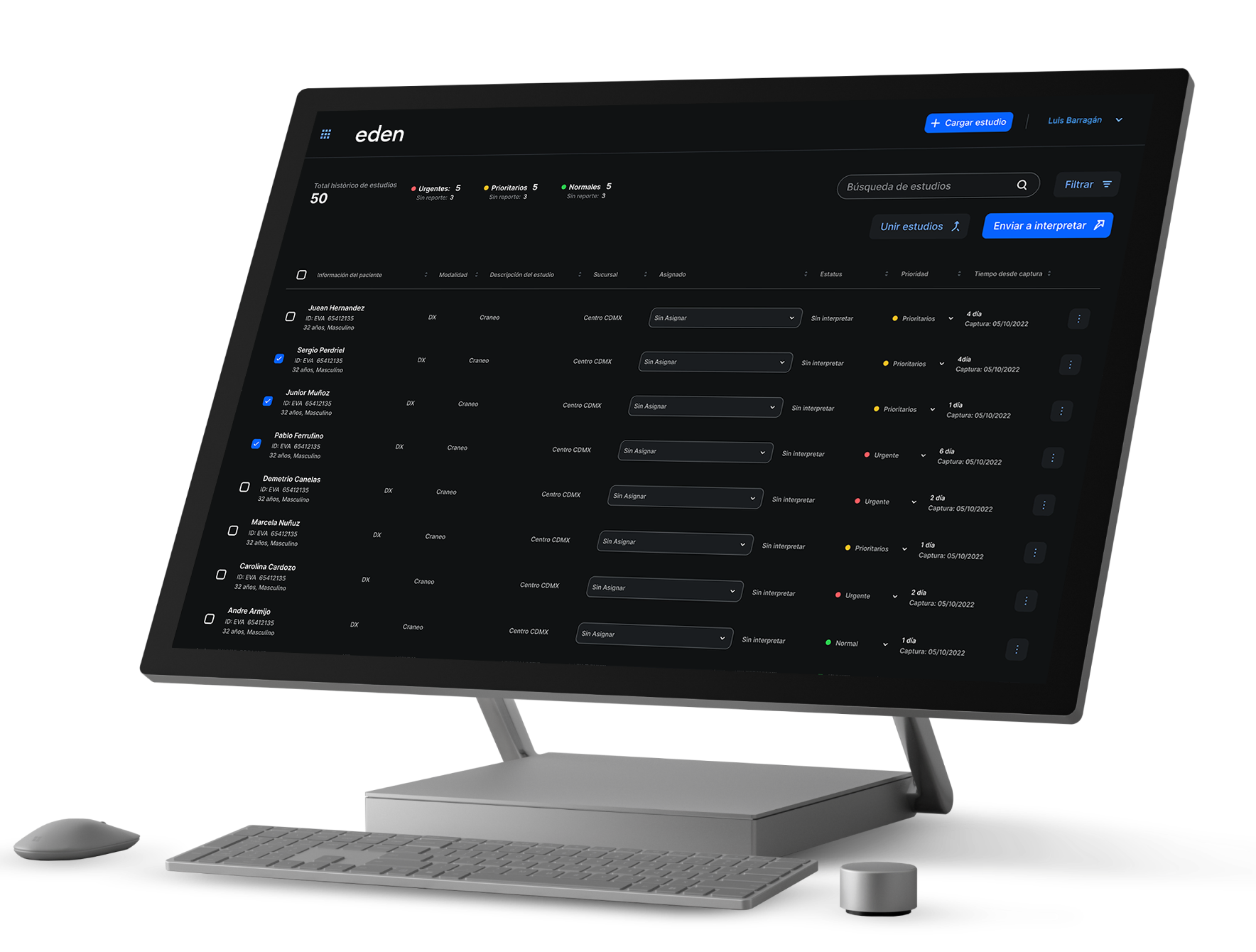


La digital radiology can be defined as computer-based X-ray technology that, instead of acetates as was done before, uses digital images. In this way, hospitals can save costs and produce quality studies that can be used for diagnostic and preventive purposes.
How does radiology work?
Traditionally the X-rays they had to be chemically processed with a method similar to that used to reveal photographs. But now, with the arrival of the digital radiology technicians use digital sensors and X-ray equipment to capture internal images. For example, it is common for dentists to use this method to take panoramas of the teeth, jaw and skull.
Direct digital radiology
When we talk about direct digital radiology we refer to sensors that send a digital image without any intermediary to a computer.
How direct digital radiology works
Direct sensing systems use a conductive material (usually selenium) which is applied on top of an array of thin-film transistors. In this way, when it comes to image transmission systems, of course, the direct radiology (DR) which use flat active matrix panels that consist of a detection layer deposited on an active matrix of thin-film transistors and photodiodes.
Features of direct digital radiology
When it comes to the way in which the patient interacts in direct digital radiology, sensitive plates are used to capture information during the study. At the same time they are transferred to a computer system.
Indirect digital radiology
Indirect systems use an X-ray intensifier screen that converts them into light. This light is then recognized by the flat detector system.
How indirect digital radiology works
In the indirect digital radiology computerized radiography systems are used (CR, for its acronym in English) which use photostimulated luminescence screens to capture the X-ray image, instead of the traditional film used previously.
Features of indirect digital radiology
To be able to observe the images from the studies, indirect digital radiology uses X-rays and an intermediate cassette.

.png)


