
Whitepapers
The latest capabilities of the Diagnostic OS


Browse all
Artificial Intelligence in Radiology: Ally or Obstacle?
Artificial Intelligence (AI) is here to stay, but we decide how to welcome it, and how to implement it. For providers of intelligent solutions like Eden, it is crucial to consider how radiologists interact with these tools in their workspaces. This can make the difference between an AI that works with the physician and one that works against them.
AI in radiology today
The field of radiology employs a variety of intelligent tools for image analysis and the generation of diagnostic impressions. These programs are capable of analyzing large amounts of information in seconds, thereby reducing the physician’s workload, speeding up emergency diagnosis, and even revealing findings previously invisible to the radiologist.
The risks of AI-related errors
But like any other technology, AI is fallible. According to a 2023 study based on knee osteoarthritis diagnoses, the error rate of an AI is generally equivalent to that of a human radiologist. However, depending on the interaction with the radiologist, these incidents can increase significantly and lead to incorrect decisions. Risk factors for medical error caused by AI include:
- Lack of understanding of how the algorithm works (also known as the “black box” effect)
- Absence of ROI or other visual AI indicators to flag its findings on the study image
- Automatic insertion of AI-generated findings into the study report
AI that keeps the physician in control
These results point to an implementation of AI in which the radiologist’s agency is not prioritized. Raymundo González, CTO at Eden, believes this should be a crucial consideration for any intelligent tool in the sector: “everything must be designed to give the physician complete control.”
Raymundo leads the development of Eden Creator, a language intelligence model that provides precise diagnostic conclusions based on the findings of a study. The goal of this tool is not to replace the physician’s interpretation, but to complement their written report, a task that “does not always require the radiologist’s skill and expertise.”
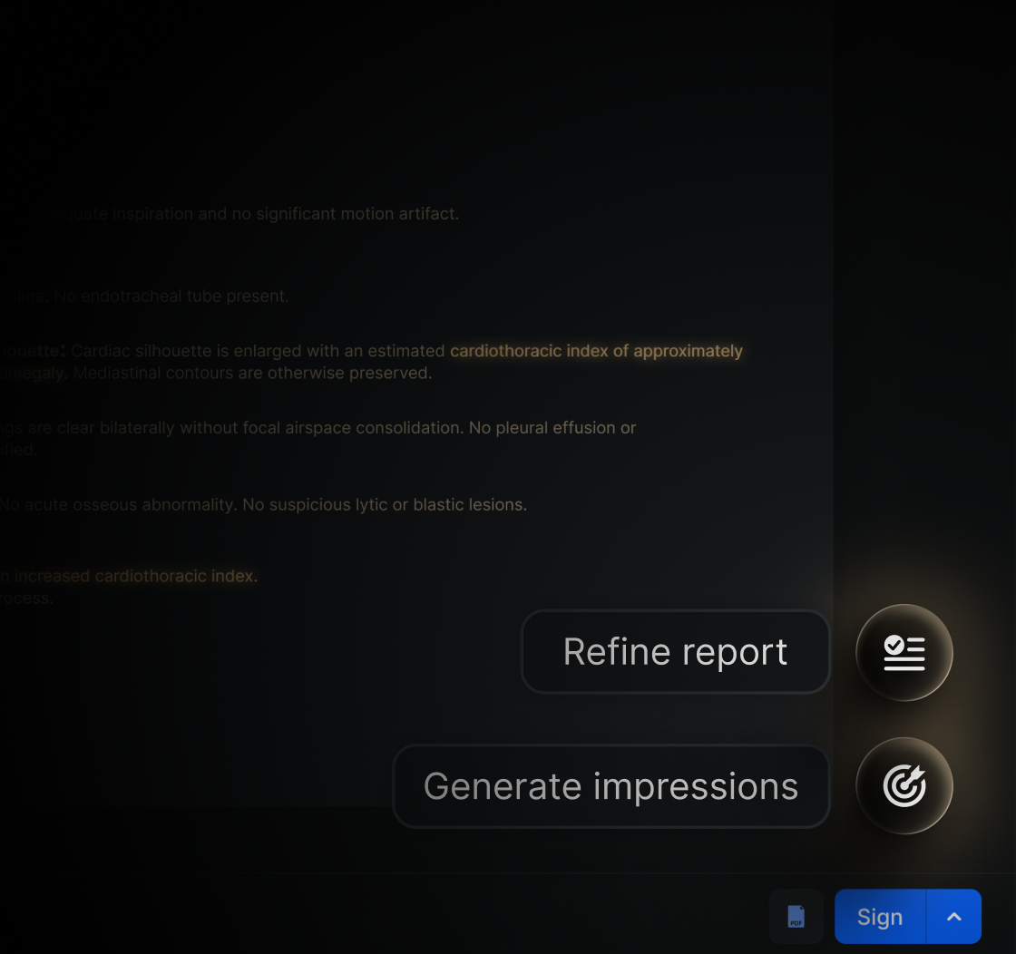
Creator is designed to empower the user, who has the ability to review the suggested conclusions, edit them, accept them, or simply reject them. With each instance, the algorithm learns from these expert decisions to refine its predictions further. This helps avoid the risk of an erroneous conclusion resulting from AI use. In addition, this tool can save the radiologist up to an hour of work, reducing the fatigue and eye strain that can also lead to medical error.
AI as the radiologist's ally
As intelligent tools become increasingly accessible worldwide, it is essential to explore implementation approaches that take the radiologist’s working conditions into account and mitigate the risk of medical error. As the American College of Radiology (ACR) states, “AI is never “an alternative” to the radiologist,” but rather an ally at their service.
References
Bernstein, M., Atalay, M. K., Dibble, E. H., Maxwell, A. W. P., Karam, A. R., Agarwal, S., Ward, R. C., Healey, T. T., & Baird, G. L. (2023). Can incorrect artificial intelligence (AI) results impact radiologists, and if so, what can we do about it? A multi-reader pilot study of lung cancer detection with chest radiography. European Radiology, 33(11). https://doi.org/10.1007/s00330-023-09747-1
Lenskjold, A., Nybing, J. D., Trampedach, C., Galsgaard, A., Brejnebøl, M. W., Raaschou, H., Rose, M. H., & Boesen, M. (2023). Should artificial intelligence have lower acceptable error rates than humans? BJR|Open, 5(1). https://doi.org/10.1259/bjro.20220053
.avif)
Humanizing Teleradiology: How to Close the Gap
Telemedicine is not the future; it is the present. Since the health emergency caused by COVID-19, remote care has become a standard tool. Despite bringing benefits such as reduced operating costs, greater accessibility for patients, and even more personalized care, telemedicine also presents challenges. The remote modality and the use of technology can turn this practice into an obstacle, diverting medical care from its true purpose: improving the patient's life.
Some professionals consider that remote medicine can be dehumanizing. This is especially concerning in teleradiology, since the physician interprets images asynchronously and rarely interacts with the patient in real time. Failing to account for the unique aspects of these situations risks producing patient distrust and limiting the quality of care they receive.
Strategies are needed to keep the patient at the center of remote radiology and to adapt current practices to new forms of digital interaction. This calls for action not only from physicians and healthcare institutions, but also from providers of medical technology systems. That is why, at Eden, we build teleradiology platforms around three fundamental pillars: autonomy, privacy, and accessibility.
Autonomy
It is crucial to prevent the patient from becoming the object of radiology, rather than its focus. Since the process of sending and interpreting images takes place independently of the patient, they may feel like “just another number” in the radiologist’s daily routine.
Effective communication is key to empowering the patient once again: strategies such as explaining the dynamics of remote interpretation in advance, offering digital follow-up through appointment reminders, and using everyday contact channels such as WhatsApp minimize patient reluctance while reaffirming the physician’s commitment to their health. Sending digital results through Eden PACS also helps build this trust, giving patients the freedom to view their medical studies from the location of their choice. In this way, individuals are able to take control of their own health.
Privacy
Sometimes uncertainty does not arise from the teleradiology process itself, but from the channels used to carry it out. Many patients are accustomed to hearing about insecure networks, hacking, and other cyber threats, which would naturally make them distrust online medical care. That is why it is essential for us that all Eden platforms comply with strict confidentiality and data protection regulations: for example, the Health Insurance Portability and Accountability Act (HIPAA), as well as corresponding certifications from the International Organization for Standardization (ISO).
Equally important is making this fact visible to reassure the patient. This includes establishing clear rules about who has access to the patient’s clinical record, continuously updating information security systems, and building strong alliances with referring physicians to create a secure digital space. This makes it possible not only to protect patient privacy, but also to give patients the freedom to provide clinical information without risk, enabling more accurate and personalized care.
Accessibility
Using accessible technology can make the difference between excellent medical care and care that excludes the patient. This is particularly true for patients with limited access to electronic devices, as well as older patients or those with disabilities, who often require support to operate digital platforms.
For this reason, every teleradiology strategy must include methods to bring patients closer to and familiarize them with new modes of care. It is vital that patient interaction platforms be user-friendly and adapted to everyday devices, such as smartphones or laptops. From scheduling appointments to delivering results, we believe technology should be accessible both economically and in terms of user experience. This principle guides the design of our user interface.
Teleradiology is a method that offers new opportunities to deliver quality medical care to patients around the world. However, to implement it correctly, it is necessary to consider the challenges it presents in terms of human interaction. Institutions, physicians, and especially telehealth technologies must place the patient at the center of their operations and establish firm regulations on bioethics, cybersecurity, and accessibility, so as to guarantee dignified medical care. Our passion is to create a secure, seamless, and friendly ecosystem that keeps the human dimension of medical service present. By making current practice more flexible and adaptable, it is possible to reduce the distance imposed by digital barriers and turn it into a catalyst for progress.
References
Cusí Sánchez, M. V., Gomes Da Costa, F., Bossio, P., Fábrega, C., García Abejas, A. and Salvador Vergés. (November 10, 2022). Telemedicine and eHealth: reflections from bioethics. Instituto Borja de Bioética (Borja Institute of Bioethics). https://www.iborjabioetica.url.edu/es/blog-de-bioetica-debat/telemedicina-y-esalud-reflexiones-desde-la-bioetica
Mesa Maldonado, M. A. (2019). The Medical Act in Light of Telemedicine: Is It Necessary to Update the Concept? Pontificia Universidad Católica de Chile (Pontifical Catholic University of Chile). UC Repository. https://doi.org/10.7764/tesisUC/MED/23664
.avif)
Eden and Apple Vision Pro: A Leap into the Future for Radiology
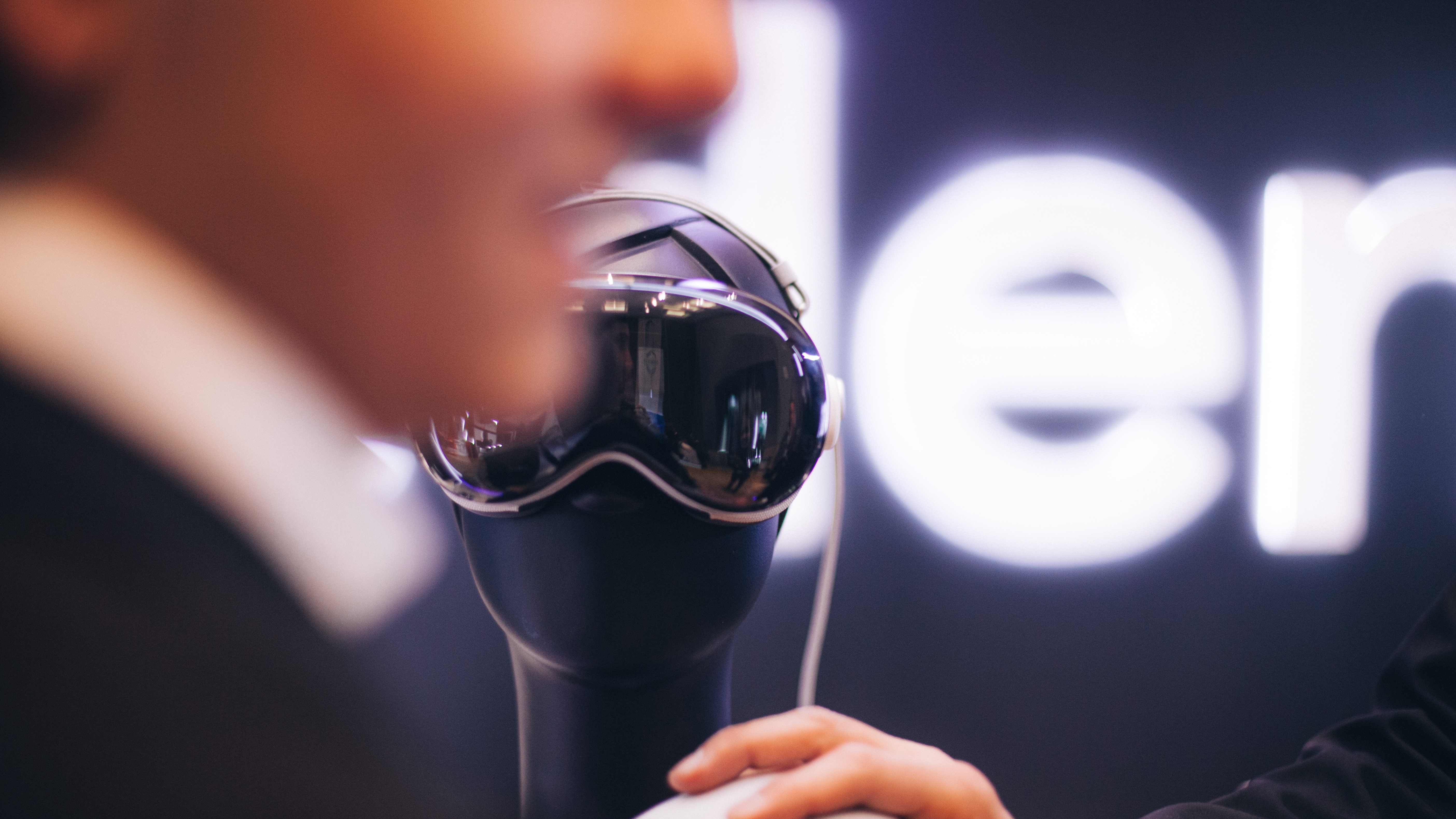
Apple's new augmented reality headset, Vision Pro, opens the door to completely transforming medicine. Eden PACS can now be used on this device to view imaging studies, marking a milestone in the use of spatial computing for radiology.
Augmented reality in radiology
Augmented reality makes it possible to project images into an immersive space. In radiology, this means an arsenal of possibilities such as navigating, manipulating, and analyzing studies. What could previously only be done through a monitor will now be possible right before the physician's eyes.
Advanced technology within everyone's reach
Until recently, this costly technology was inaccessible to the average radiologist. Now, Vision Pro puts it at everyone's service, with portable headsets. This is especially important in LATAM, where there are only 30 radiologists per million people, and many lack the appropriate equipment to do their job.
In this context, Vision Pro represents a transformative shift that creates opportunities to bring advanced technology to thousands of radiologists at a lower cost, increasing productivity and improving the quality of healthcare across the continent.
As Eden's CEO, Julián Ríos, states: "now any physician will be able to have 4K monitors, in whatever size they want, right at their fingertips, directly on their head, in their eyes."
.jpeg)
What it means for physicians and patients
In the very near future, this accessible technology will be crucial for making the best decisions for patients' benefit: physicians will be able to collaborate remotely to handle emergency cases in an instant, imaging students will be able to train with realistic, high-definition cases, and interventional radiology will have more precise tools to minimize risk during delicate procedures. In addition, thanks to its eye-tracking technology, users will be able to view and manipulate studies naturally, thereby reducing the burnout that afflicts the vast majority of radiologists worldwide.
"This is not a hypothesis, it is not something that might happen in the future. It is something that is already possible: today, our users, if they have Vision Pro, can access Eden PACS. Doing so is a quantum leap in what radiology can do today for physicians and patients."
Eden PACS and the democratization of radiology
Eden PACS exists to drive the democratization of advanced radiology throughout LATAM. We provide more than 600 clients in 16 countries with a cloud-based radiology solution and instant digital delivery, with no additional installation costs. We also put vital tools in radiologists' hands, such as MPR reconstruction, specialized suites for orthopedics and mammography, and even artificial intelligence for report generation. Augmented reality will make it possible to bring this technology to more people and elevate radiology in the developing world to new heights.
Our sacred purpose is to ensure extraordinary healthcare for everyone. With Vision Pro, we make this vision a reality.

RIS System: What Is a RIS System and What Is It For?
A radiology information system is software used to manage medical images and other associated data in a hospital setting. Discover its main features and benefits.
Technology is becoming increasingly incorporated into the healthcare sector, making digitization more prevalent. This has benefited both the physician-patient relationship and all the functions of the hospital facility. Notable examples are RIS systems. Let's take a look at what they consist of.
What is a RIS system?
The term RIS stands for radiology information system (radiology information system). It is responsible for collecting, controlling, and exploring all the data obtained from the radiology diagnostic service.
A RIS collects all the information on a patient's radiology activity, from the moment the study is ordered until the results report is completed. This entire system is designed to be easy to use.
With a RIS system, it is possible to identify everyone involved in performing the diagnostic test. This allows each person to be held accountable in the event of an error, as well as improving the service provided. It also helps the head of the radiology and imaging department with decision-making.
RIS systems generally operate using worklists, which help increase productivity and provide greater control and organization. The essential worklists of a RIS are:
- Appointment waiting list.
- Exam worklist.
- Diagnostic worklist.
- Transcription worklist.
- Supervision worklist.
Functions of a RIS system
Some of the main functions offered by a RIS system are:
- Appointments. This is the assignment of a date and time for the study, which can be manual or automated.
- Reception. Manages the processes involved when the patient arrives to undergo the test.
- Activity log. A record containing information on the radiology technician, matches, time taken, and materials used.
- Reports. The creation of the radiology report, which is later sent to the patient or the referring physician.
- Statistics. Involves data mining within the system to obtain important information, such as workload.
- Billing. Allows tracking of payments for services rendered.
- PACS integration. Allows control over the storage of radiology studies and other functions specific to a PACS system.
Characteristics of a RIS
Among the important characteristics that make up a RIS, we can mention:
- Image storage, tracking, and distribution. A RIS system stores each radiology image, along with its corresponding report and all the data pertaining to the patient. It also allows tracking of how all information is shared through the computer network connected to the RIS (whether local software or cloud storage), so that physicians can access the data whenever they need it.
- Patient and resource management. By opting for this digitized service, everything is kept in an organized record that is easy to access for authorized staff. This makes patient registration, scheduling, and material management much easier.
- Patient tracking. Allows verification of all patient information, medical history, studies performed, current treatment, and progress by logging into the system.
- Interactive documents, input and output of results. The digitization of all documents helps improve the distribution of information, reducing wait times, improving communication among physicians, and achieving a faster and more accurate diagnosis. All results are reported digitally and can easily be sent by email or instant messaging. There is also the option to print when needed.
PACS-RIS integration
During the interpretation process, prior studies or reports are frequently needed and must be available immediately upon request. This means that certain preparations must be made in advance, and this process must be automatic. When a study or scheduling order is placed, if the RIS sends information about the scheduled study, the PACS can automatically retrieve or preload relevant prior studies.
If a study is changed immediately before it is performed, or if it is added as an emergency, the new information can be sent to the PACS for further review. At any point during interpretation, the radiologist should be able to request and view a patient's previous reports from the PACS workstation.
It is important to know that a RIS system complements a PACS system very well. Each provides its own functions and tools, which brings stability to the radiology and imaging department, and therefore to the entire hospital.
Preparing in advance for the installation and adoption of a RIS is key to success. To minimize the risk of failure, it is important to follow these steps:
- Seek specialized IT consulting.
- Plan an incremental migration.
- Ensure adequate hardware and communications infrastructure.
- Ensure the acquisition of a RIS that implements HL7 and certifications such as HIPAA.
- Anticipate future systems to be integrated with the RIS, such as the PACS system.
- Review the department's workflow and reorganize it if necessary.
- Train and familiarize staff with the information systems.
Advantages of the RIS system
Implementing a RIS system brings numerous advantages, among which we can mention:
- Reduced costs for support and handling operations.
- Losses in the storage of radiology film are avoided.
- Service delivery is improved, since once the image is captured, the result is immediately transmitted to the PACS and the image can be viewed in real time.
- It is environmentally friendly, since film production is avoided and paperwork is likewise reduced.
- The diagnostic process is improved, since data entry errors are significantly reduced.
- Priority is given to physician-patient care.
- Increases patient flow due to more efficient appointment scheduling and faster reports.
References
- The Future of the Radiology Information System. American Journal of Roentgenology. Vol. 200, No. 5.
- C. Martínez Serrano: Sistemas de Información Radiológica. Sistemas de información radiológica.
- Janice C. Honeyman. Information Systems Integration in Radiology. Journal of Digital Imaging, Vol 112, No 2, Suppl (May), 1999.

HIS System: What Is a HIS System and What Is It For?
Hospital facilities manage a large amount of information that, in most cases, used to be scattered and not immediately available. For this reason, systems have been developed to help manage this information in a way that is useful for decision-making, such as the HIS system. Find out what it is used for and what its main features are below.
A hospital information system, or HIS (Hospital Information System), is one designed to meet the needs of a hospital facility, such as storing, processing, and reinterpreting medical-administrative data. All of this allows for the optimization of human and material resources.
Every HIS system will generate reports depending on the hospital area for which it is required, providing feedback on the quality of healthcare service delivery. It is important to note that its use relies on security measures, generally based on standards such as HIPAA (Health Insurance Portability and Accountability Act), which ensure the authentication, authorization, and management of user profiles for those who interact with the available data.
Within a hospital, there are several types of information systems. HIS integrates them to achieve the automation of many kinds of transactions and processes. It is important to consider the existence of major system groups, such as:
- Front office. These are systems that support the delivery of healthcare services, focused on the institution's external interactions.
- Back office. These support the functioning of the hospital's departments, focused on the institution's internal operations.
- Clinical systems, diagnostic and treatment support. These automatically gather the patient's clinical information and data.
In addition to these, there are several specialized services within a hospital, including: clinical laboratory, pathology, pharmacy, etc. Each of these has information systems that have been optimized to function according to the specific requirements of each area.
Requirements for implementing a HIS system
To implement a HIS system, certain basic elements are required, which will vary depending on the service provider.
- Intranet and internet communications network.
- Hardware equipment, depending on infrastructure and financial resources.
- Base software with which the system will be programmed and operated.
- Staff training for using the system and its information.
- Financial funding.
Types of HIS systems
According to their intended functions within the hospital system, HIS systems can be classified into these types:
- Administrative HIS. Manages all hospital resources, including human, material, and financial resources.
- Medical-administrative HIS. Manages the relationship with patients and the data they generate as users of hospital services.
- Clinical HIS. Refers to the patient's state of health or illness, as reflected in the medical record.
Functions of a HIS system
The main function of a HIS is to manage information regarding a hospital's services and make it accessible to all authorized staff.
Likewise, a HIS is responsible for providing support for activities at various operational and tactical levels within a hospital. For this reason, computers are used to gather, store, process, and communicate clinical-administrative information.
Other functions of HIS systems include:
- Keeping control of all services provided to patients.
- Obtaining general statistics on patients.
- Obtaining epidemiological data.
- Detailing the cost of care provided to each patient.
- Maintaining a strict electronic medical record.
- Providing patients with access to information in a timely and appropriate manner.
- Giving them the ability to update that information and exercise their right to correct data.
- Improving clinical practice through decision support.
- Supporting teaching and research activities.
- Making the institution's specific plans more efficient.
- Harmonizing scientific-technical information with administrative-accounting information.
Advantages of HIS systems
Once a HIS system has been implemented, the clinic or hospital will benefit in the following ways:
- Retrieval of information on patients and hospital processes is done more effectively, since everything is automated.
- A greater volume of information can be handled while still keeping up with daily tasks.
- All information can be managed in real time.
- Reduced paper use, since recorded information is digitized.
- Better monitoring of supplies, inventory, and medications.
- Ensures the availability of information for decision-making.
- Minimizes response time to patients, clients, and suppliers.
Traditionally, HIS systems can be integrated with other medical systems designed to increase productivity, generate savings, and improve patient care, such as PACS and RIS systems.
References
- Conceptos generales HIS. Oficina de inteligencia sanitaria.
- Sistema de Información Hospitalaria. Sistema Mid.
- Leonardo Yunda Perlaza y Luis Fernando Gómez. Informática Médica: Sistemas de Información y Estándares en Salud: Modelo de Aplicación.

Essential Tools of a PACS
A PACS has become an extremely useful tool for medical centers with a radiology department. These systems allow physicians to review all files related to a patient's studies digitally.
Digitization of medical documents
The main goal of these systems is to allow medical images to be moved and managed more quickly, so that medical and administrative staff, as well as patients themselves, benefit.
PACS components
Now, when it comes to choosing the PACS that can be put to the best use, there are several aspects to consider. The first is to assess the medical center's objective, as well as its specific needs. However, when it comes to PACS components, there are some that definitely cannot be missing. Below we list five of them.
DICOM viewer
It is true that there are countless different image formats; however, in order to standardize medical studies, the DICOM format, Digital Imaging and Communications in Medicine, was created. This guarantees that all physicians can correctly view this type of image and know it is the standard format to use for any study, whether CT scan, MRI, ultrasound, etc. In addition, this type of image is also accepted in other systems with RIS or HIS, in addition to PACS.
Measurements in a PACS system
There are different types of measurements that can be relevant when interpreting a study, such as distances, angles, ROI, annotations, among others. With a PACS such as Eva, the radiologist can take these measurements while they are automatically saved in the toolbar that shares the same name. Other measurements that can be important include the cardiothoracic index and the Cobb angle.
Cloud storage of medical images
Radiologists face heavy workloads, so it is not uncommon to find them working at several medical centers. But what happens if, for some reason, they cannot travel to their workplace? With a cloud-hosted PACS, the physician can receive studies and do their work from wherever they are, with the assurance that the patient will receive their diagnosis promptly.
Cine mode in medical PACS
When dealing with very long image series, it can be useful to view them in motion, as well as to choose the number of frames per second for playback. Eva's development team created this tool with precisely this need in mind. It also makes it possible to see details that cannot be observed in a static view.
Custom tools for radiology images
There are many tools that can be added to PACS systems to make physicians' work easier. At Eva Center, our team strives to create new shortcuts every day and add whatever our partners need. Along these lines, we offer voice dictation or recognition, as well as 3D reconstruction, which can be extremely useful for a better viewing perspective, multiplanar reconstruction, and many others.

How to Measure the Usability of a PACS System
The relationship between medicine and technology is becoming increasingly relevant due to the simplicity with which certain procedures can now be performed compared to how they were carried out a few years ago. For this reason, there is a key concept for measuring how viable a given tool is: usability. More and more attention is being paid to how user-friendly a given program or website is.
In the case of PACS systems, where images and reports from various studies are stored electronically, it is of great importance that they include the necessary options for medical staff to perform an appropriate interpretation. In the following section, we will describe what a PACS system is and how it works.
What does a PACS system consist of?
As mentioned earlier, PACS systems store information, mainly images from radiology studies. Previously, film printing was used, so these systems have come to replace that task. The main difference is that these PACS systems feature structures that allow for greater quality and precision.
Usability of a PACS system
There are different criteria for determining the usability of PACS systems. However, some of the most important aspects to consider are the number of available tools, the way they are laid out, and the average number of clicks it takes to complete an interpretation. There are also other factors, such as automatic sequencing and the ability to customize the system. The goal of all this is to increase the productivity of medical staff.
On the other hand, the learning curve is also relevant, since the more intuitive the PACS is, the less time it will take a physician to become familiar with the tool.
Once an institution acquires a PACS system, at some point a new physician will arrive who will also need to use this tool. That same professional is likely to work at different institutions and therefore use PACS solutions from different vendors, which is why having user-friendly tools will ensure they can do their job better.
Another relevant aspect of usability is whether the physician constantly needs an IT professional to make configuration changes. If the answer is yes, this means that usability is inadequate. This applies even more if a workstation connected to a server is used, since in that case the physician must be in that same location to interpret studies, whereas with a cloud-based PACS, only a computer with internet access is needed. If we add to this the difficulty of requiring a technology expert, the PACS's usability drops.
PACS components
A PACS has various components, such as modalities for imaging equipment, communication networks, archiving systems, workstations, and software. For example, if a physician is diagnosing a mammogram, they can choose, depending on what they need, to use two quadrants, one for the right breast and one for the left. If they want to compare it with a study from six months earlier, the screen could be divided into four quadrants. The degree of usability will depend on how simple it is to bring up these options.
It can also define the image slice thickness, use cine mode to view it in motion, or display it in 3D
So, if you are looking to switch to or acquire a new PACS, you now know some of the most important elements to evaluate in terms of usability, a fundamental recipe for these tools that will undoubtedly bring great benefits to both physicians and patients.

Hospital Infrastructure Management
As the renowned American philosopher Ralph Waldo Emerson once said: "The first wealth is health," a statement society identifies with to a great extent, since people daily seek out the best hospital services for any symptom, control process, or prevention effort. Hence, hospital infrastructure management must stay at the forefront of existing advances to maintain and increase patient trust.
Now, when we talk about hospital infrastructure, it includes several elements, such as:
- Internal building structure
- Interior design
- Lighting
- Parking
- Green areas
- Access zones
- Equipment
- Information systems
In other words, this infrastructure relates to the building itself, the resources for treating patients, and also the comfort experienced at every moment within the hospital. Indeed, even the colors and decoration of the rooms, though it may not seem so, are important for conveying calm and confidence in the facility's level of professionalism.
How can hospital infrastructure be improved?
Many hospitals stand out in their category due to their expertise in different specialties, a fact that makes it considerably more difficult to fully remodel their infrastructure. However, when a facility is well regarded by patients, continually improving the most critical points delivers optimal results in the medium term.
To begin with, it is necessary to conduct a study of the aspects that cause the most discomfort for patients, sometimes parking, decoration, or the turnaround time for radiology studies, to name a few examples. Based on this, hospital management can then determine where to begin investing.
To identify patients' pain points when visiting the facility, a fairly straightforward approach is a simple multiple-choice survey. It is worth noting that these studies statistically reveal dissatisfaction with turnaround times, which is why adopting technology that reduces patient wait times is key to improving hospital infrastructure.
The importance of hospital infrastructure
As mentioned in the previous sections, patient satisfaction is what maintains or increases the profitability of healthcare facilities. In that sense, having all the elements that make them up be state of the art guarantees success.
If we talk about the building and its decoration, people naturally associate them with being able to find the solution they need to stay healthy, of course, guided by experts with extensive experience in specific specialties.
Likewise, a good parking area is appropriate so that people have easy access. Sometimes hospitals lack this service, and as a result, some people rule out the facility, even knowing that the best doctors are there.
As for technology, it is a highly valuable asset for patients, since society is increasingly immersed in it. Therefore, finding applicable digital solutions in hospitals, such as PACS systems, is appropriate for attracting more patients.
Infrastructure of a top-tier hospital
The most notable aspects of a top-tier hospital's infrastructure are:
- Building and decoration. Modern construction featuring bright, uplifting colors such as white is the leading trend in world-class hospitals. These are linked to patients' self-esteem.
- Green areas. Some regulatory considerations must be taken into account here, since certain plants and trees attract wildlife that can affect hygiene and health aspects. Therefore, the type of flora and its placement must be carefully evaluated, though nature undoubtedly appeals to patients.
- Access areas. Doors and parking spaces are essential for people with mobility limitations, making them a crucial part of hospitals.
- Technology. State-of-the-art equipment and systems that streamline hospital operations are essential today. PACS (Picture Archiving and Communication System) systems are one such example, as they enable the capture, storage, and transmission of radiology images in record time among hospitals, specialists, and patients.

What Is Ultrasound and What Is Ultrasound Used For?
Medical technology has succeeded in harnessing different methods to determine what is happening inside our bodies. Such is the case with ultrasound, or sonography, one of the most common radiology and imaging studies, which physicians across many specialties may order for the diagnosis and treatment of numerous diseases or injuries. Below we explain how it works.
What Is Ultrasound?
There are many types of waves, and one of them is mechanical waves, which are used in ultrasound. These are sound waves not perceptible to the human ear, which can be used to diagnose various conditions, as well as to assess the general health of our bodies. In gynecology, ultrasounds are commonly used to track the progress of a possible pregnancy or the health of the reproductive system.
Ultrasound also has therapeutic applications following injuries, mainly orthopedic injuries, which may involve immobilization.
Ultrasound is nothing more than the conversion of electrical energy into mechanical waves, and this is done by the medical equipment also called an ultrasound or sonography machine. It is a noninvasive process, and therefore neither painful nor harmful to the body. This is why there is no limit to how many ultrasounds, or other studies based on this same principle, you can undergo.
These mechanical vibrations occur upon contact with an elastic medium, such as the skin. The sound waves bounce off the structures of our body by means of a transducer, which in turn displays a real-time image on a monitor.
How Does Medical Ultrasound Work?
In medical ultrasound, waves are used to diagnose many conditions or diseases associated with gynecology and the digestive system, although it is also used in cardiology, urology, orthopedics, ophthalmology, endocrinology, and other medical specialties.
The most important part of the equipment used for diagnostic ultrasound is the transducer probe, since it is responsible for sending and receiving waves to create the echo effect that produces images of the body. A cold gel is applied to the area of the body being examined, which helps transmit the body's images.
Another use of ultrasound is in various surgical procedures, using sterile probes to assess the internal structure to be treated as the procedure progresses. Such is the case with ultrasound-guided biopsies, which give the interventional physician a clear picture of where to take the tissue sample that will be examined under a microscope.
This type of ultrasound, in turn, has two categories: the first is the anatomical, which is used to obtain images of the body's internal structure, whether tissues or organs. The second is the functional, which allows the velocity of tissues or blood to be merged with other information to produce “information maps.”
One of the most important advances in ultrasound is that results can now be obtained in 3D, 4D, and color. This latter feature is known as Doppler ultrasound. All of this has been extremely helpful for studying the heart and its proper function, as well as arteries, liver inflammation, and many other regions of the body.
Ultrasound for Therapies
When we refer to therapeutic ultrasound, there is no change in the underlying principle of generating mechanical waves from electrical energy; the only difference is that in this case, the specialist will not give you images as results.
The goal is for these waves to interact with the tissues of your body in order to modify them according to the pre-existing condition. Some examples include breaking down blood clots and tumors, heating certain areas to relax the muscle, and directing medication to specific locations.
This makes it possible to reduce inflammation and pain; it increases skin permeability without leaving any associated scarring, boosts enzyme production, and offers many other advantages. Depending on the purpose, there is an established frequency dose and a technique that can be either continuous or pulsed.
Are There Risks Associated with Ultrasound?
Ultrasound is noninvasive and painless for your body, which is why experts in medicine and radiology consider the risk associated with these studies and therapies to be nonexistent, especially compared with the radiation exposure involved in X-rays.
However, you should know that these are two completely different studies with specific purposes depending on the area of the body to be assessed for a subsequent diagnosis, which is why only your physician can determine which imaging study is best for you.
To ensure your body's safety, what matters most is that you visit certified healthcare centers with extensive experience, ones that stay at the forefront in terms of equipment and updated information.
References
- Ultrasound. Terapia Física.
- Ultrasound. National Institute of Biomedical Imaging and Bioengineering.

What Is Computed Tomography and What Is It Used For?
Below we will explain what computed tomography is and what its main applications are. This radiology and imaging study is one of the most frequently ordered by experts to make their diagnoses.
One of our main wishes is to have health and well-being, and for this to be possible, it is essential that whenever we experience any symptom or trauma, we see a specialist physician, who will tell us the next steps to take. While we do indeed need to wait for these instructions, it is important to know that our physician may very well request imaging studies in order to make a diagnosis.
This is because symptoms alone, or a simple description of what happened, are not enough for the physician to determine the extent of any condition affecting our body, whether due to an illness, or after suffering an accident, blow, or fall. So do not be surprised if, when you visit the specialist, they ask you to undergo radiology and imaging studies, since this is one of the most reliable methods for examining the internal structure of the human body.
What Is Computed Tomography and What Is It Used For?
One of the most common imaging studies is computed tomography, or computed axial tomography (CT or CAT scan), which involves two techniques for obtaining detailed images of a body area of interest. The first is the use of X-rays through a tube that rotates around you, allowing captures to be taken from different directions. The second is computer technology.
Computer technology is what allows the physician to obtain more detailed and sophisticated images of the body. In addition, this technique allows for computer manipulation to more precisely view small areas of the body, such as blood vessels, the bronchi, sections of the spinal cord, and tissues, among others.
It should be noted that the advantages mentioned above give computed tomography scans added value and therefore a higher price compared with plain radiographs (X-rays). In addition, although the process is painless, it takes a bit longer (around 30 minutes).
The above is due to the fact that the technologist or radiologist must follow a set of steps to ensure a quality tomography image, such as proper calibration of the equipment. If this is not done correctly, the study may not turn out well and you would have to undergo the radiation again. That is why we suggest going to a quality laboratory, clinic, or hospital.
Contrast Media in Computed Tomography
In certain cases, and to achieve better visualization of the inside of the body, the patient may be instructed to receive a contrast medium. This can be administered orally or intravenously.
These contrast agents are widely used and are not risky. However, you should tell whoever is performing the study if you feel any symptom of excessive heat or pressure when it is administered, as this could indicate an allergy. Afterward, it is advisable to drink plenty of fluids to help flush it out.
The facility where you have the study performed will give you the appropriate instructions for preparation before and after the study if a contrast agent has been indicated.
What Can a Physician See With a CT Scan?
Since CT scans produce sophisticated images that can be professionally manipulated on a computer to get a better view of your body's internal structure, their scope is considerably broad.
This is why a physician may order one to evaluate any of the following diseases or medical conditions:
- Investigating possible internal bleeding, for example after cardiovascular damage.
- Assessing internal injuries or fractures of both the skeletal and muscular systems.
- Locating tumors, clots, or infections. On this last point, we can mention pulmonary conditions such as pneumonia, in which contrast agents are commonly used to highlight the condition.
- Guiding surgical procedures to determine where to make incisions. A simple example is biopsies.
- Monitoring conditions such as cancer or pulmonary diseases.
Is Getting a CT Scan Dangerous?
Indeed, when you undergo a CT scan, your body receives radiation that is higher than what you are usually exposed to naturally. However, technological advances have allowed radiologists to control radiation doses in order to reduce the associated risks.
These doses depend on the area being scanned. For example, for the abdomen and pelvis, there is an exposure of 10mSv, equivalent to two and a half years of natural background radiation. For the lungs and chest, the figure is 1.5mSv, corresponding to 6 months; and for the teeth, at 0.18 mSv, it is similar to 22 days of radiation.
How Do I Prepare for a CT Scan?
In general, no prior or special preparation is required. In some cases, you may be asked to fast or stop taking fluids or medications beforehand, but this usually occurs only when contrast media are used.
It is suggested that you wear comfortable clothing. You may be asked to remove your clothes and put on a medical gown. Likewise, you will not be allowed to wear any metal accessories of any kind.
Notify the physician if you suspect you might be pregnant, or if you are pregnant, before undergoing the study.
When it is time to begin, you will simply need to lie down on a table. The study will be finished within 10 to 30 minutes.
How Will I Get the Results of My CT Scan?
The results are generally accompanied by an interpretation from the radiologist. Keep in mind that only your physician knows what each term means, and it is up to them to decide what steps to take regarding your treatment.
You will generally receive your results between one and three days after your study is performed. Many laboratories and hospitals allow you to view them digitally. That way, you can save them and easily share them with additional physicians, for example if you need a second opinion.
References
- Computed Tomography. MedlinePlus.
- X-Rays, CT Scans, and MRIs. OrthoInfo.
- CT Scan. Mayo Clinic.
- Radiation Risks from Imaging Tests. American Cancer Society.

Why Is It Important to Have Quality Technical Support for a PACS System?
Learn about the benefits of having technical support for a PACS system, and why quality service also translates into long-term savings for any healthcare institution.
Today, technology is advancing by leaps and bounds, both in our everyday lives and in various fields of knowledge, among which healthcare systems clearly stand out. In this sector, technology facilitates patient care and makes work in hospital organizations more efficient.
One example of this is PACS systems, because in addition to being secure and increasing the productivity of operations, they make work easier and provide easy access to information.
However, for the entire system to function properly, there must be certainty that if something goes wrong, a well-prepared, efficient, and available technical support service will be there. A powerful infrastructure is of little use if, when technical support is needed, requests simply go unanswered.
That is why we will now discuss the advantages and benefits of quality technical support for healthcare technology solutions, such as PACS systems.
Characteristics of a Quality Technical Support System from a PACS Provider
When choosing a PACS system, it is very important to ask the provider about the responsiveness of their technical support.
Likewise, if a PACS system is already in place, it is worth evaluating whether quality technical support is included. No one wants to wait until a crisis occurs to find out whether there will be someone available to help resolve the problem.
Some of the factors of a quality technical support system that can be taken into consideration are the following:
- The main aspect is accessibility, since information must always be available to the medical staff who require or need it. Technical support must ensure that all staff have access to and know how the systems work, even before the PACS is implemented at an institution.
- In this regard, a quality PACS system must provide training not only at the start of implementation, but on an ongoing basis. This training should be provided whenever there is new staff at the healthcare institution, whenever there are system updates, and so on.
- Another important factor is security. The PACS system must guarantee that security measures are in place, and in the event of a breach, technical support must ensure a prompt and efficient resolution. It should also provide data backup and the implementation of security measures and standards, such as HIPAA.
- Without a doubt, another essential factor is availability. Quality technical support will offer fast response times, as well as different channels of communication. Likewise, technical support must be available around the clock, especially if the healthcare service operates 24 hours a day or handles emergencies.
- Network management. Acquiring a PACS system facilitates communication over computer networks and enables the transmission of images to different remote locations within the hospital through the communication network. But what happens if this communication fails or is disrupted? Efficient technical support must be able to resolve any issue in this regard, since the hospital or clinic cannot afford service interruptions.
- Support for image processing. With a PACS system, images are processed using different methods in synergy with the diagnostic modalities. Quality technical support must know how to make the appropriate adjustments so that all modalities can be used seamlessly with the PACS system.
- Price. When we acquire any technology service, we assume that technical support is guaranteed, but this is not always the case with PACS providers. When choosing a PACS system, questions should be asked about service, maintenance, licensing, and update costs. A quality service will include personalized attention and technical support within the cost of the system itself.
Why Choose a PACS System?
Today it has been shown that PACS systems offer great capacity and speed, facilitating better diagnosis and effective management of hospital information.
In the not-too-distant future, PACS can be expected to become a fundamental piece within the framework made up of the various information systems present in a modern healthcare institution.
Among other elements, PACS systems offer the following benefits:
- Today's healthcare organizations come to form complex networks of affiliated institutions that provide comprehensive patient care services.
- As for the efficient exchange of information among the various areas and sectors related to the patient, a communication system is needed that fully incorporates all clinical data, which means that diagnostic images are an important and fundamental component.
- Areas explicitly dedicated to the healthcare sector need to be able to compete by lowering costs without sacrificing service efficiency, and to keep innovating for the benefit of the quality of care provided to patients. All of this is possible through the implementation of PACS systems.
- In addition, having a PACS system is considered a highly competitive tool and a strategic investment for improving both productivity and the organization's reputation within the hospital environment.
All of these factors depend directly on the PACS system provider being able to back its services with personalized attention and technical support that is efficient, reliable, and available at all times.
References
- Hernández, Raúl. “Desarrollo de habilidades en el uso de las tecnologías de la información y la comunicación. PACS” (Developing Skills in the Use of Information and Communication Technologies. PACS).
- Silva Alvines, Eduardo. “Introducción a sistemas RIS-PACS” (Introduction to RIS-PACS Systems).
- “Sistemas para archivo y comunicación de imágenes (PACS)” (Picture Archiving and Communication Systems). Guía tecnológica no. 41. Secretaría de Salud (Ministry of Health), México, 2009.

T1 and T2 Magnetic Resonance Imaging: TR and TE Weighted Images
Many physicians feel apprehensive about not being able to interpret this terminology since they are not trained as radiologists. To help prepare you for when you encounter these terms, we will show you how to gain a better understanding of this useful tool in the medical field so you can provide better healthcare.
What Is Magnetic Resonance Imaging and How Does It Work?
Magnetic resonance imaging (MRI) is a diagnostic method used to produce detailed images of organs and tissues by means of a magnetic field and radio waves that change rapidly. The resulting images are then displayed on a computer to determine whether a lesion is present and to what extent.
Magnetic resonance imaging is an interaction between an external magnetic field, radiofrequency waves, and atomic nuclei. When a body is subjected to a magnetic field and subsequently stimulated by electromagnetic waves (radiofrequency waves), the result is resonance of its atomic nuclei.
The basis for obtaining images is measuring the energy released and the time it takes to return to a relaxed state once stimulation ceases. Through this radiological study, high-quality images of the human body are obtained, enabling accurate diagnoses. Thanks to magnetic resonance imaging, we have been able to reveal the different cross-sections of tissues, organs, and vascular structures.
Basic Radiology Concepts in Magnetic Resonance Imaging
Setting aside purely technical aspects, what is important to understand is the sequential process:
1. The hydrogen nuclei are the key players in forming the image by MRI. When the patient is placed in the MR scanner, the hydrogen atoms, previously oriented at random, align with the static magnetic field.
2. To detect the signal, a radiofrequency pulse is briefly applied, producing a net change in the alignment of these atomic nuclei.
3. When the radiofrequency pulse ceases, the spins return to their equilibrium state, releasing energy to the surrounding molecules.
4. The rate of energy release is determined by the intrinsic relaxation properties of each tissue, characterized by the longitudinal relaxation time (T1) and the transverse relaxation time (T2).
5. T1 represents the recovery of longitudinal magnetization in the direction of the main magnetic field.
6. T2 represents the loss of magnetization in the transverse plane, perpendicular to the axis of the field.
7. Substances with a long T1 (for example, fluids) will appear dark on T1-weighted images, while those with a short T1 (fatty tissues) will show a high-intensity signal.
8. On T2-weighted images, a substance with a long T2 (fluid) will appear bright.
9. The main advantages of MRI are its excellent contrast resolution, high spatial resolution, and the absence of ionizing radiation.
10. Among the agents approved for clinical use in MRI, the most commonly used contrast agent is a drug that shortens T1, called gadopentetate dimeglumine (or Gd-DTPA), because it contains gadolinium, a paramagnetic agent (atoms with unpaired electrons in their outer shells).
11. Tissue relaxation is altered by the interaction between gadolinium's unpaired electron and tissue hydrogen protons, which significantly shortens the T1 of blood relative to the surrounding tissues.
12. MR images can be obtained using different “sequences.” The most commonly used are known as spin-echo (or spin echo) sequences and can be weighted in either T1 or T2.
T1-, T2-, TR-, and TE-Weighted Images
MRI allows healthcare professionals to detect abnormalities and lesions that help confirm a diagnostic hypothesis and improve treatment guidelines. This is why we must deepen our understanding of T1-, T2-, TR-, and TE-weighted imaging.
Many physicians fear they will be unable to interpret this terminology since they are not trained as radiologists. Below we show you how to gain a better understanding of this useful tool in the medical field and its most common terms.
To more precisely understand what relates to a weighted image, we must recall the basic planes of every magnetic resonance study, understanding that it works with three planes: the axial, coronal, and sagittal planes.
When it comes to gaining a deeper understanding of the qualities of this imaging study, in this case magnetic resonance imaging, it is important to be familiar with certain concepts described previously in order to understand what is meant by a weighted image.
The pixel value and contrast of an image are determined by intrinsic and extrinsic factors:
- Intrinsic factors: proton density, T1, T2, flow, and diffusion.
- Extrinsic factors: TE, TR, flip angle, TI (inversion time), turbo factor, echo train length, b-value, among others.
Definition of Key Terms in Radiology
- T1-weighted images. This is the measure of relaxation time as it manifests in the longitudinal plane. These images are created primarily using data from the relaxation rates that differentiate protons in the longitudinal plane of a main magnetic field.
- T2-weighted images. This refers to the measure of relaxation time as it manifests in the transverse plane. These images are created using the same principle as T1-weighted imaging, except that it occurs in the transverse plane.
- TR-weighted images. Describes the measure of repetition time. In other words, it is the time needed to complete a full excitation/relaxation cycle for a pulse sequence, expressed in milliseconds (ms).
- TE-weighted images. This is the measure of echo time. That is, it is the time elapsed between the start of a pulse sequence and the acquisition of data from excited protons, expressed in milliseconds (ms).
What Is a Pulse Sequence?
The echo time (TE) and the repetition time (TR) refer to a concept known as the pulse sequence, a series of instructions repeated numerous times, which allows data to accumulate in order to ultimately create an MRI image.
The pulse sequence has two essential variants:
- Spin echo pulse sequence. This event begins with a 90-degree pulse and continues with a 180-degree pulse, generating protons that create the MRI signal. This type of sequence is used to weight images in T1 and T2.
- Fast spin echo (FSE) pulse sequence. The advantage of this type of technique is that, after the 90-degree and 180-degree pulses, multiple echoes can be obtained. In other words, multiple slices of the scan are obtained. This modality is much faster than conventional spin echo, especially for T2, since it requires a shorter repetition time (TR) to create the image.
Difference Between a T1-Weighted and T2-Weighted Image Based on TE and TR
Generally speaking, a T1-weighted image has a short TR and TE, the former being less than 1,000 ms and the latter less than 20 ms. On the other hand, substances with a long T1 will be hypodense, while a short T1 will result in a substance that is hyperdense in terms of intensity.
A T2-weighted image has a long TR and TE, the former being more than 2,000 ms and the latter more than 40 ms. In this case, substances with a long T2 will be hyperdense, while hypodense substances will remain hypodense.
How to Distinguish a T1-Weighted from a T2-Weighted Image?
- On T2-weighted images, fluid is hyperintense, whereas on T1 it has an intermediate signal.
- Fat is brighter on T1, although it is not the ideal predictor. On T2 it is hypointense.
- On T1, hyperintense structures include blood, proteinaceous substances, melanin, and paramagnetic agents (gadolinium).
- On T1/T2, structures such as air, cortical bone, ligaments, tendons, fibrous tissue, and blood flow appear hypointense.
On a T1-weighted image, the bone marrow of a normal adult (fatty or yellow marrow) has high signal, that is, it is hyperintense or hyperdense. This means it appears white in tone, while cerebrospinal fluid has low signal, that is, it is hypointense or hypodense, giving it a dark tone.
Nervous tissue, such as the spinal cord or nerve roots, has an intermediate signal intensity. Cortical bone, which lacks mobile protons to produce a signal, is hypointense on all pulse sequences.
On T2-weighted images, bone marrow has lower signal intensity, cerebrospinal fluid becomes hyperintense, and nervous tissue maintains an intermediate signal intensity. However, the spinal cord has relatively lower signal intensity, since it is surrounded by cerebrospinal fluid, which has a much higher signal intensity.
The intervertebral discs of normal individuals typically have an intermediate signal intensity on T1-weighted images and, due to their water content, appear hyperintense on T2-weighted images.
Diagnostic Value of T1- and T2-Weighted Images
One of the main uses of a T1-weighted image lies in its application in neurology, since the image quality and the precision of anatomical detail allow for a more accurate diagnosis. It also helps determine the best therapeutic course for the patient.
The advantage of a T2-weighted image is that it shows fat as a low-intensity signal (hypo) and fluid as a high-intensity signal (hyper). This, for example, is very useful in lesions and pathologies characterized by an increase in fluid content. There is also a reversal with respect to white and gray matter, with white matter showing lower intensity.
The mechanism used by radiology and imaging equipment may seem complex, but understanding how an image is obtained allows us to improve patient diagnosis. Let us remember that the type of contrast used is key when performing a paraclinical study.
Today, this type of procedure is enhanced thanks to the technology used by PACS systems (Picture Archiving and Communication System), which give specialists the opportunity to improve image contrast and quality using a variety of techniques and tools.
References
- E. Scott Pretorius and Jeffrey A. Solomon. 2006. Radiology Secrets. Elsevier.
- Joaquín Costa Subias and Juan Alfonso Soria Jerez. 2015. Resonancia magnética dirigida a técnicos superiores en imagen para el diagnóstico (Magnetic Resonance Imaging for Senior Diagnostic Imaging Technicians). Elsevier.
- Michael Y. M. Chen, MD, Thomas L. Pope Jr., MD, and David J. Ott, MD. 2006. Radiología básica (Basic Radiology). Mcgraw-hill - Interamericana de España.
- “Resonancia magnética” .Temas especiales. Manual MSD versión para profesionales.
- “Cómo interpretar las Imágenes de Resonancia Magnética”. Radiología 2.0.
No papers match your search.