
Whitepapers
The latest capabilities of the Diagnostic OS


Browse all

ROI DICOM: What Is the ROI Tool in a DICOM Viewer?
What Is the ROI Tool and What Is It Used For?
This is a tool whose English acronym stands for “Region of Interest,” that is, region of interest. It refers to a part of the image that one wishes to filter or operate on in some way, for example to make an annotation or measurement.
In a DICOM viewer, the ROI is one of the basic functions. Different PACS systems offer different ways to mark the region of interest, or ROI, on a study, for example circles, ellipses, polygons, and rectangles, among others.
Through the properties of the viewer and the tool itself, both its appearance and its behavior can be customized.
Its main use is to perform measurements once the region of interest, or ROI, has been detected in an area of the medical image. This allows the physician to make comparisons with previous studies, adjust contrast or brightness, take measurements, and generally use all the tools of the DICOM viewer around the desired area.
Other tools of the DICOM viewer that most commonly interact once a region of interest, or ROI, has been detected include:
- Length measurement
- Angle measurement
- Area measurement
- Volume measurement
- Annotations
- Polygonal area selection
- Freehand area selection
What Is DICOM?
An acronym for Digital Imaging and Communication in Medicine, DICOM is a standard for digital imaging and communication in medicine. It was designed to specify a protocol of rules for data exchange, the digital image format of medical studies, and a file structure for medical images and associated related information exchanged between imaging equipment from different vendors.
DICOM was originally developed by the National Electrical Manufacturers Association (NEMA) and the American College of Radiology for computed axial tomography (CT) and magnetic resonance imaging (MRI) scans. It is currently maintained by the DICOM Standards Committee and supports a wide range of medical images in the fields of radiology, cardiology, pathology, and dentistry.
DICOM is used in virtually every modern hospital. It ensures the interoperability of the systems used to produce, monitor, transmit, query, process, retrieve, and print medical images and derived structured documents, as well as to manage clinical workflow through a PACS system.
How Does the ROI Relate to the DICOM Viewer?
Generally, diagnostic modalities such as magnetic resonance imaging, computed tomography, and computed radiography, among others, can be costly in terms of both image storage and infrastructure. For this reason, a way was sought to compress these images to make them more manageable for both storage and distribution, without affecting their resolution or diagnostic capability.
The ROI tool is also a form of compression, such that the area of the image belonging to the region of interest can be compressed using a lossless algorithm in order to recover the original data without alteration. Likewise, the outer area is compressed using a lossy, or irreversible, algorithm, which can likewise achieve results with a visually indistinguishable reduction in quality.
The DICOM protocol introduces the ROI tool through the capabilities of the JPEG 2000 image standard, allowing the area of greatest diagnostic importance within the image to be compressed losslessly, while the remaining region is compressed with moderate loss.
The ROI tool can be specified through a set of coordinates or as a binary mask the same size as the image.
There are three alternatives for ROI-based encoding:
- Tiling mechanisms
- CodeBlock Selection
- Coefficient Scaling (referred to as the Maxshift algorithm in the JPEG 2000 standard)
Among the functionalities of the DICOM protocol are extracting images in various formats, parsing the header, and packaging files into new DICOM files, as it is capable of writing headers that comply with the standard.
The ROI, in turn, must define a region of interest based on the interpretation of a set of coordinates or an image in PGM (portable graymap) format. The resulting ROI is returned in a standard format using JPEG 2000 compression. It accepts different input image formats and produces a sequence of output images with the (.jp2) extension. Its capabilities include the ability to apply lossy or lossless compression and interact with the ROI component to use the region-of-interest technique during compression.
References
- DICOM general description Overview.
- ROI-Based Processing. Math Works.
- González, L. “An Approach to Medical Image Compression Based on Regions of Interest and Motion Compensation”. Congreso Argentino de Informática y Salud (Argentine Congress on Informatics and Health), CAIS 2013

Impact of Digital Transformation in Mexican Hospitals
With the world now fully immersed in a digital environment, and with so many technologies at our disposal, the healthcare system has found ways to innovate and develop solutions where they were needed. Learn how this has been achieved in Mexican hospitals.
Digitalization has become extremely important in today's modern world, and a sector as significant as healthcare and medicine could not be left behind. Digital transformation in hospitals must focus on improving patient care, facilitating physicians' work, reducing human error, and cutting costs.
2020 was identified as the year with the highest global investment in health technologies worldwide. In the context of the pandemic and everything happening at the healthcare level, investments in health digitalization tripled compared to previous years.
For the benefit of the healthcare system and patients, digital systems have now been incorporated into Mexican hospitals. Mexico's healthcare system is made up of several important entities, including physicians, patients, and private and public hospitals. All of them have advanced in this shift toward digitalization.
What Is Health Digitalization?
Health digitalization involves the use of technologies to eliminate the constraints of time and space, as well as extending the reach of the healthcare system to wherever patients are located. This makes it possible to achieve four important objectives:
- Achieving better outcomes and increasing treatment quality through access to the right professional regardless of where the patient and specialist are located.
- Improving access and convenience for patients, eliminating the need to travel or relocate when it is not possible or convenient to see a specialist, which improves the patient experience.
- Improving physicians' experience by reducing the need to travel. This allows them to focus on patient care throughout their career and on more suitable workflows.
- Cost reduction by leveraging scarce physicians and specialists across multiple clinical sites, hospitals, and offices simultaneously.
In other words, health digitalization helps provide specialized care and reduce the likelihood of medical errors. The use of technology makes it possible to integrate evidence-based best practices into professionals' clinical workflow, ensuring, for example, that the right medications are administered at the correct dose, at the right time.
Many errors can be avoided by using the electronic health record, which is fundamentally important. The same applies to clearly defined and documented care plans, structured assessment tools, and clinical decision support for physicians.
On the other hand, PACS systems make a significant contribution to the digitalization of medical services, and also enable teleradiology.
Benefits of Technology in the Healthcare System
- Clinical records that are clearer, more accurate, and more accessible, staying continuously updated as healthcare providers collect data on their patients.
- Critical processes are simplified, saving time whenever a patient has to fill out a form, since all information is linked and available in the event of an emergency.
- Higher-quality data is collected, since it carries a lower margin of error than manual data entry.
Digitalization in Mexican Hospitals
According to one study, by 2016 health digitalization services had been installed in 30 hospitals in Mexico City, with the patient as the top priority.
The digital clinical record is the first step toward securely managing each patient's data. As a result, interoperability is achieved among these hospital centers, improving patient safety and continuity of care. Likewise, greater service efficiency is achieved along with cost reduction from avoiding repeated tests.
Some of the benefits obtained through this implementation included:
- Optimizing care processes and reducing patient wait times.
- Increasing prescription safety through the integration of standardized medication databases and the creation of drug alerts.
- Improving the informed consent management process (biometric signature).
- Improving the quality of health data through the implementation of the digital clinical record.
- Increasing patient safety and diagnostic accuracy.
Due to the pandemic, the use of available technologies had to be accelerated to avoid overwhelming Mexican hospitals. Both physicians and patients had to grow accustomed to using telemedicine as a way of accessing healthcare, starting with technological tools such as the phone, through video calls or virtual conferencing tools.
At the public health level, the Instituto Nacional de Enfermedades Respiratorias, Ismael Cosío Villegas (National Institute of Respiratory Diseases, INER) is Mexico's leading research center for respiratory diseases, as well as a highly specialized hospital in the field. With the support of Cisco, a global IT company, it installed a series of applications to improve service and open up new medical areas. This, in turn, provided the connectivity needed to implement remote visits, teleconsultation, streamlined medical reporting, and physician-patient interaction.
Impact on Mexican Society
The digitalization of Mexican hospitals has had a positive impact on society. With remote care, while preserving the quality of the physician-patient relationship, wait times are reduced without the need for exposure or travel. A mobile phone and an internet connection are all that is needed to establish highly effective communication.
However, there is an economic barrier, since not all Mexicans have access to a mobile device, computer, or internet connection, which makes it difficult to reach every patient.
This is according to statistical data presented in the 2020 Encuesta Nacional sobre Disponibilidad y Uso de Tecnologías de la Información en los Hogares (National Survey on Availability and Use of Information Technologies in Households, ENDUTIH), which found that only 78.3% of the urban population uses the internet. Among the rural population, usage stands at 50.4%.
As for remote medical consultations, they account for 16% of private practice in Mexico, according to a study on the digital habits of the medical community. It was also found that 84% of physicians are connected online, mainly via smartphone. The main place of connection is the institution where they work (46%), followed by home (29%), which means continued investment is needed to consolidate this practice.
Without a doubt, it remains a priority to keep investing in the shift toward digitalization of healthcare institutions in Mexico. Although significant investments have been made in infrastructure, more is still needed to consolidate these changes.
References
- Digitizing and connecting healthcare to improve efficiency and quality of care with ehCOS technology SAMIH_2016_v4 Success Story.
- Press Release. National Survey on Availability and Use of Information Technologies in Households, 2016. Inegi.
- 1st Study on Physicians' Internet Habits in Mexico, April 2021. IRP.

Plate Digitizer: What Is It and What Is It For?
Discover how you can digitize physical radiological plates to store them in the cloud. This is especially useful if you are considering migrating your practice to a PACS system.
Today, many technological advances have emerged that have improved and corrected small errors or complications in many aspects of the healthcare sector.
Previously, in order to make diagnoses from the radiological images produced on plates, physicians had to view and analyze the image in analog form. Today, with all the advances available, diagnoses can be provided using nothing more than a computer and an internet connection through PACS systems.
What Devices Exist for Digitizing Physical Plates?
If your medical practice uses physical plates, but you are considering digitizing your system, there is an option that will allow you to back up studies digitally and in the cloud. One way to achieve this is with a plate digitizer.
A plate digitizer, also known as an X-ray digitizer, is a piece of equipment that converts your analog plate into a digital format in a matter of seconds so you can view and edit it on your computer.
Are Plate Digitizers Expensive?
X-ray digitizers can seem costly when purchasing the equipment. However, when thinking ahead, you will discover all the time and materials you can save over the equipment's service life.
The prices can vary, as with any product you might purchase. Prices also depend on the product's range and brand. A professional, medical-grade piece of equipment can cost between 10 and 14 thousand dollars. However, other manufacturers offer much more affordable options. Below, we discuss other alternatives for digitization.
Are They Easy to Obtain and Operate?
This equipment is not easy to find, in the sense that you will not find it in a shopping mall or store. You will most likely need to contact a manufacturer or supplier to request a quote and, if necessary, import the equipment.
Since these are digital, versatile, and not very complex devices, they will not be very complicated to use.
What Types of Radiological Plate Digitization Exist?
Digitization consists of transforming the original information from the analog or physical plate into a digital composition and format that will make it possible to view, process, and manipulate all the image data on a computer.
One type of plate digitizer develops the study without the need for conventional developers or darkrooms, which makes it possible to save a great deal of time and reduce development costs.
On the other hand, when acquiring a PACS system, images are automatically converted into digital format using the DICOM protocol, the global standard for medical images. The most sophisticated and comprehensive PACS systems are compatible with all diagnostic modalities and perform this digitization step automatically.
Another advantage of being able to store all physical plates in a PACS system is that you can keep them in the cloud, as an extra layer of protection against data loss from theft, misplacement, or damage to computer equipment.
Radiographic plates can also be digitized using a CCD camera, which offers adjustment of the black-and-white and gray values of the image through an optical system.This camera, known as a charge-coupled device (its English acronym is CCD, charge-coupled device), uses an internal circuit for image digitization.
Finally, there are specialized medical scanners that operate with an optical system that measures the image point by point. In this way, a resolution superior to human vision is achieved.
How Are Images Archived in the PACS System?
The acronym PACS stands for Picture Archiving and Communication System. These are systems that enable the management of all medical information, improving the productivity of any medical organization.
Generally, the PACS archive consists of a short-term and long-term storage system.
- Short-term storage. This consists of a set of identical disks (RAID) that provide very fast access to image data.
- Long-term storage. This consists of magnetic tapes or optical discs.
It works as follows: after 3 to 30 days, the image is moved from RAID to long-term storage. To access that information, it must be copied back to short-term storage.
Today, the cost of RAID storage has decreased thanks to cloud systems such as Eva PACS. This is because, being linked and stored in the cloud, they only use short-term storage (RAID), thereby eliminating the need to retrieve the long-term archive. This has also brought advantages in terms of security.
Why Were There Complications with Printed Plates in the Past?
In the past, there were various shortcomings when performing a radiology study in relation to image-based diagnoses. Such complications were associated with the following:
- Difficulty comparing studies.
- Slowness in accessing all the patient's information.
- No database record or history was kept.
- Exams had to be repeated due to lost plates or errors in the original studies.
These were the main problems a physician could encounter when making a diagnosis, resulting in substandard patient care.
Looking at today's advances, we find that with systems such as Eva PACS, there is not even a need for a special application, since the system can be accessed directly from a web browser. For this reason, with Eva PACS it is also possible to digitize all your physical plates and keep them in a very secure place using only your computer, saving you time and reducing costs.
References
- Information and Digitization: Centennial Congress of the Sociedad Argentina de Pediatría (Argentine Society of Pediatrics)
- Radiographic Digitization, Josep Alfred Piera i Pelliçer.
- Radiology Secrets, E. Scott Pretorius & Jeffrey A. Salomon.

Computed Tomography: Image Manipulation
Imaging studies have become the most important tool for assessing patients' conditions and making more reliable diagnoses, which can be reviewed with other specialists through teleradiology. For these results to be of high quality, however, image manipulation techniques also play a role, for example, in computed tomography (CT).
In tomography, images can be sectioned into parts using computer systems to obtain an image with a broader scope of analysis. These manipulations are image acquisition techniques. Below, we will look at some of them to provide patients with more accurate answers.
Preliminary Considerations for Quality Tomography
To begin using a scanner, certain steps must be followed, such as turning on the CT equipment, then connecting the Gantry console, which houses the X-ray tubes, the information panels, the radiation detection devices, and which activates the structure. The tubes must also be warmed up according to the manufacturer's manual, and finally, calibration must be performed.
This calibration is performed daily or monthly according to the provider's specifications. In this way, the software avoids errors to achieve quality, optimized images, as established by various image quality standards. This allows specialists to verify that the parameters and slices are correctly configured.
Additionally, to access image manipulation tools, it is necessary to have a PACS system whose DICOM viewer includes them.
Techniques for Image Manipulation in CT
Below, we will review some of the most widely used image manipulation techniques in interpreting CT scans. All of these techniques are made possible by the advanced tools of the DICOM viewers found in the best PACS systems.
Recall that image manipulation techniques are the techniques or processes that modify a radiological image to improve the visibility of useful information, while reducing information that is not useful for diagnosis, sometimes known as noise.
Multiplanar Reconstruction (MPR)
First, we will discuss multiplanar reconstruction (MPR), one of the most widely used techniques and one that offers the greatest benefits for accurate diagnoses.
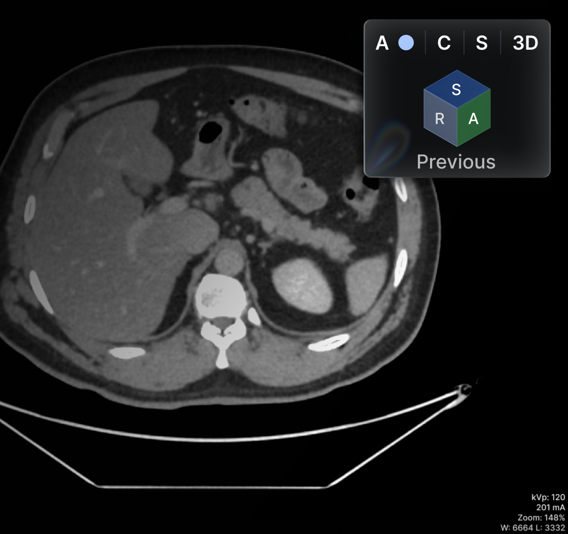
This technique makes it possible to go from a two-dimensional (2D) image to a three-dimensional (3D) plane in order to view cross-sections of the body area in question in axial, sagittal, and coronal planes.
The dimensional change in MPR is built from a single series or image acquisition, and there are no alterations in its quality. In effect, to be able to use this technique, the healthcare facility must have a cutting-edge technological architecture, like the DICOM viewers of the most sophisticated PACS systems.
Maximum or Minimum Intensity Projection (Mip/Minip)
Another manipulation technique is maximum or minimum intensity projection (Mip/Minip), which is useful for zooming in on and highlighting very small body structures that are therefore difficult to evaluate. Examples include blood vessels, specific tissues, bronchi, and areas to which contrast was applied for more precise observation.
Some experts consider the Mip/Minip technique to be a zoom tool, but in reality, knowing how to section the part to be evaluated requires skill in order to make the necessary close-ups, especially when dealing with small body structures that are difficult to visualize.
Hounsfield Scale (Measurement Tool)
For its part, the Hounsfield scale makes it possible to highlight the outline of the body area to be evaluated and to take measurements of the region. All of this is useful for differentiating the flow of blood, water, and other substances, which can likewise be highlighted with contrast.
Being able to take measurements and adjust the scale using the Hounsfield principle depends on the temperature and pressure of the attenuation or projection of the X-ray beams. Therefore, resolution can be enhanced in areas of the body where there was greater radiation absorption.
The Relationship Between PACS Systems and CT Manipulation Techniques
As mentioned earlier, for all image manipulation techniques to be possible, healthcare facilities need to have appropriate technological infrastructure. This is where PACS systems come in, which must offer viewing and reconstruction options for these studies.
This once again reinforces the importance of digitalization in the healthcare sector for the benefit of both physicians and patients. Additionally, PACS systems offer many other benefits, such as patient clinical history, task visualization, worklists, and much more.
It should be noted that for these PACS systems to function properly, they need to be built on a cutting-edge technological infrastructure. In other words, a platform in which images can be processed smoothly while maintaining quality across the various slices and dimensional changes.
Recall that CT studies can contain around 125 images, weighing approximately 20 MB. Hence, the PACS provider's servers must have sufficient speed and capacity so that images can be accessed and manipulated with ease. Likewise, it is best for the healthcare facility to have access to a broadband internet connection.
Contact an Eva agent for advice on the best solution for your practice.
References
- González, C., et al. Computed Tomography and Ultrasound Techniques. Arán.
- Zúñiga, J. (2009). Picture Archiving and Communication Systems (PACS). Mexico, Secretaría de Salud (Ministry of Health).
- What Is Cloud Architecture?. Red Hat.

Teleradiology: What Is Teleradiology?
Advances in technology have contributed to the development of medicine, not only in terms of devices or medications, but also in the transfer of information. Telemedicine refers to the delivery of health services and information through electronic means. For example, it enables remote collaboration among specialists, physician education, and in some cases, patient care.
Thanks to the internet, telemedicine has been established across all medical specialties, and teleradiology has undergone significant development.
Teleradiology makes it possible to obtain medical images and store them electronically so they can be shared and transmitted remotely for interpretation, whether for diagnostic purposes or consultation with a radiologist.
This way of working is being increasingly implemented in hospitals, clinics, and medical practices across various specialties.
How Does Teleradiology Work?
The teleradiology process rests on three fundamental pillars:
- Image storage.
- Transmission network.
- Image reception.
In this regard, specialized software programs have been developed to facilitate the sending of radiological images, simplifying the process as much as possible.
Today, digitized images are regularly transmitted at high speed. PACS systems facilitate and speed up this process, allowing specialist physicians to collaborate, request second opinions, or quickly confirm diagnoses.
Benefits of Teleradiology: Advantages and Disadvantages
The greatest benefit of teleradiology is providing the patient with better care, since it allows them to receive a digital report and learn their diagnosis remotely.
It also allows specialist physicians to digitally evaluate a patient's images and prepare a diagnosis regardless of distance. This facilitates collaboration among specialists, no matter where they reside.
This translates into time savings for both parties, since it allows information to be sent without needing to travel to the place where the study was performed.
Thanks to the storage of patient information, this data can be used for future evaluations, as well as for follow-up and comparison. Patient information is safeguarded in the cloud with security and reliability within a PACS system.
Finally, teleradiology also contributes to the environment, since phosphor plates are highly polluting due to their slow degradation process, which usually takes several decades or more.
1. The advantages of teleradiology include allowing radiologists to remotely access patient images and reports from anywhere. This allows them to review patient images and reports in real time, without having to be physically present at the hospital or clinic. This can be especially beneficial for patients who need to see a specialist but live in another city or country. With teleradiology, they do not have to travel if they can be examined by a specialist from anywhere.
2. The disadvantages of teleradiology are that, although it is possible to review images from anywhere, their quality will vary depending on the internet connection available, and reviewing images may take longer because they need to load each time they are opened.
The Need for Teleradiology in the Medical Industry
Teleradiology emerged partly due to the imbalance between demand and availability of diagnostic services.
The high demand for radiology services in places such as the United States, the United Kingdom, and Singapore could often not be met by the number of in-house professionals available.
This reality is also experienced in Latin American countries, where there are few radiologists.
Delays in delivering an imaging study report, as well as the lack of available professionals, can negatively affect the patient, depending on the severity of their condition.
Therefore, the immediate availability of medical diagnostic services is extremely important, especially during medical emergencies.
Another reason that justifies the growing need to implement teleradiology is geography and the ability to provide services remotely. Many rural areas around the world lack both radiology services and the necessary personnel.
For this reason, with the help of this branch of telemedicine, this shortfall can be fully overcome through the remote services that more experienced personnel at healthcare facilities in cities can provide.
Even in cities, difficult cases in specific areas of radiology can be shared in search of a second opinion. This even makes it possible to consult other experts quickly and easily.
What Services Does Teleradiology Provide?
The term "teleradiology" refers to the process of interpreting radiological images remotely. Teleradiology uses a combination of technologies, including the internet, automated image capture, retrieval and transmission, and secure cloud storage.
The advantages of teleradiology include greater accessibility and efficiency, better image quality unaffected by physical distance or bandwidth constraints, and shorter turnaround times.
Teleradiology is used for a variety of purposes, including the following:
1. Providing remote diagnostic consultations
2. Facilitating collaboration among radiologists in different locations
3. Reducing workload and providing relief during peak hours or high-volume periods
4. Facilitating access to specialists in areas where they are limited or unavailable
5. Allowing more efficient use of resources, such as costly equipment, experienced staff, and specialists' time.
What Does Telemedicine Mean?
Telemedicine is the process of providing healthcare remotely using audio, video, or other electronic means.
Telemedicine can be used to provide services to hard-to-reach patients or those with unusual conditions. It can also be used in the care of patients who require specialized knowledge.
One of the main benefits of telemedicine is improved access to healthcare services for people living in remote areas. It can also be an effective way to care for patients with chronic conditions who do not require periodic doctor visits.
The use of telemedicine requires adequate infrastructure, including reliable internet connections and the necessary equipment
Recent Expansion of Teleradiology
Medical imaging is an essential pillar for efficient patient care and diagnosis. In recent years, it has been possible to optimize the performance of software and PACS systems.
In this regard, public and private institutions that have decided to make use of teleradiology ensure diagnostic effectiveness and patient safety, as well as avoiding additional costs for printing plates, saving time, and gaining many other benefits.
References

Multiplanar Reconstruction: What Are the Advantages?
Computed tomography scans (CT) are among the most accurate studies in radiology and medical imaging, as they allow for multiple volumetric approaches depending on the area of the body to be evaluated. The most common and efficient case is multiplanar reconstruction, known by its acronym MPR, which makes it possible to view axial, sagittal, and coronal cross-sections from the same series.
The above means that with a flat or 2D image, a slice edit can be performed to thicken the area of interest. In fact, it is practically possible to obtain a three-dimensional view starting from two coordinates. In certain studies, this technique is very useful because there are small areas that are not easily seen in the general scan.
This technological solution for reconstructing images by cross-section is truly remarkable, and its main advantage is the ability to reach more accurate diagnoses. This is achieved especially with Computed Axial Tomography (CAT), since it allows numerous planes to be generated from a single one to detail organs, tissues, and bones with greater precision. These cross-sectional views feed the coronal, oblique, sagittal planes, and 3D tools of the MPR.
It should be noted that all these reconstructions are made possible thanks to the modifications and updates that the most sophisticated PACS systems and DICOM viewers have undergone, which allow the MPR tool to be used with high quality and without any errors.
Advantages of CAT and MPR
The main advantage of CAT is that it allows multiplanar reconstructions with various types of cross-sections in a very short time. All of this without affecting the resolution of the series or image, while making room for 3D planes. This is why, for radiologists, the MPR tool becomes an indispensable ally for accurate diagnoses.
CAT results would not be as efficient and useful if multiplanar reconstruction options did not exist.
Finally, it should be added that MPR is so efficient because of isotropic voxels, or equal slice thickness and resolution, since its results are based on the computer's spatial interpretation.
Voxels are small three-dimensional subunits, usually measuring 200 µm or less, and they offer the ability to visualize the object under study with an accurate representation.
Planes Used in MPR
Recall that multiplanar reconstruction makes it possible to create images in different planes from a single original one, offering a clear visualization advantage over traditional CT planes, which only allow viewing one slice at a time. MPR creates isotropic images in which image quality is preserved at the same level as the original axial plane.
The planes that can be viewed with the MPR tool are:
- Axial. This plane crosses the body from front to back and divides it into upper and lower sections.
- Coronal. This is the plane that crosses the body from left to right and divides it into anterior and posterior sections.
- Sagittal. In this plane, the body is crossed from front to back, dividing it into left and right sections.
- Oblique. Finally, this plane crosses the body at axial, coronal, and sagittal angles.
How Does MPR Work?
This tool is based on the research of mathematician J.H. Radon, who proposed, at the beginning of the 20th century, a computational model for deriving three-dimensional data from two-dimensional data. Its application in medicine came much later.
Multiplanar reconstruction manages to graphically represent the characteristics of a two-dimensional layer, processing the data as a single set to ultimately create the three-dimensional image. The computer treats the object of study as being made up of equilateral rectangular prisms, called isotropic voxels (slices), which are the volume units used to form a three-dimensional matrix.
When viewing a 3D image using the MPR tool, what is actually being observed is a reconstruction achieved through a complex calculation, which allows tissues to be analyzed from the various desired planes. This makes it possible to calculate lengths, angles, areas, and volumes with great precision, which is useful for accurate diagnoses of various conditions, something impossible with other imaging techniques.
Sophisticated software with an MPR tool will make it possible to adjust contrast and enhance desired aspects of the area of interest.
PACS Systems and MPR
All these multiplanar reconstructions derived from CT and CAT scans are made possible by the installation of a tangible and intangible technological architecture. This requires computers with cutting-edge software, such as PACS system DICOM viewers, to display imaging studies of different modalities, such as computed tomography.
When choosing a PACS system, it is important that the provider offers security and reliability in the exchange of information. For this to happen, these platforms must also operate according to imaging (DICOM) and text (HL7) standards, so that interoperability, productivity, and communication take place optimally, as is the case with Eva PACS.
It should not be forgotten that "reconstruction speed depends on the power of the computer and also on the reconstruction algorithm used" [Rottke, Dennis]. Eva PACS offers a fast multiplanar reconstruction tool, or MPR, which will provide more faithful and accurate diagnoses, as well as help in developing an effective treatment plan.
We interviewed Dr. Benjamín Conde Castro, a physician specializing in Radiology and subspecializing in Oncological Imaging and Breast Imaging. Here is Dr. Conde's opinion on the DICOM viewer of Eva PACS, specifically regarding the multiplanar reconstruction feature:
"I think any radiologist would prefer speed over tools that might slow the system down. I always prefer speed, and I think it's the same for any radiologist. For example, I like how fast Eva's MPR tool works."
References
- What Are MPR, MIP, MiniP, and Volume Rendering Reconstructions? Sociedad Española de Imagen Cardíaca (Spanish Society of Cardiac Imaging).
- SLD253 Multiplanar Reconstruction Module for Computed Axial Tomography Studies. Universidad de las Ciencias, Cuba (University of Sciences, Cuba).
- Computed Tomography: Introduction to Dental Applications. Scielo.
- MPR. WikiRadiography.
- Rottke, Dennis. "Primary and Secondary Reconstruction in Digital Volumetric Tomography". Quintessence, Vol. 23, No. 9.

X-rays: What Are They and What Are X-rays Used For?
Radiology and medical imaging studies are increasingly used in healthcare services to diagnose different diseases and injuries. Countless clinical studies are performed on patients to detect fractures, tumors, infections, deformities, degeneration, and conditions in the body's various systems. X-rays are indispensable due to their great usefulness, practicality, and low cost.
What Are X-rays?
You may be wondering what X-rays are. Put simply, we can define them as a type of radiation that uses beams of rays or electromagnetic waves to project the internal structure of the body. Performing X-ray studies is essential in cases where laboratory tests or physical examinations are not sufficient, since they allow the physician to see what is happening inside our body.
Some of the studies in which X-rays are used include X-ray imaging, mammography, computed tomography, and dental panoramic radiographs. Although each is performed differently using specific techniques, all of these studies can show the bones, tissues, and organs in the area under study.
When this study is performed, X-rays are absorbed by the desired parts of the body, revealing their internal structure based on the amount of radiation received. Depending on this, different shades will appear. For example, bones absorb the most waves due to the calcium in the skeletal system, appearing as a white tone. Meanwhile, tissue, muscle, and fat interact less and appear as gray shadows.
Imaging Studies That Use X-rays
X-ray Imaging
The electromagnetic waves emitted by X-rays are not visible to the naked eye, but several studies use them to explore the internal structure of the body. X-ray imaging is the best known, and is used to evaluate different conditions, for example trauma (impacts), conditions such as osteoporosis and arthritis, infections and metabolic disorders, cancer, and foreign objects in soft tissue.
This test is fast, painless, and efficient, since no preparation is required beforehand. It is only important for women to inform the radiologist if they are pregnant, so that they can consult with the treating physician on whether exposure to radiation is advisable.
Computed Tomography
There are other imaging techniques that make use of this penetrating energy to study the body. Such is the case of computed tomography (CT). Physicians request it mainly for more detailed analysis, because of its unique ability to transform 2D images into 3D, showing bones, organs, tissues, and more with greater clarity.
Angiography is a type of tomography in which specialists seek an image of the veins and vascular system. A common application is that it allows visualization of the structures through which blood enters and exits the heart.
On the other hand, there is multislice computed tomography (MSCT), which captures the scans in layers. It is also widely requested in cardiology.
Mastography or Mammography
Finally, we can mention mammograms, X-ray images of the mammary glands that use X-rays to explore their structure and detect any abnormality. It is one of the preferred methods for the early detection of breast cancer.
Breast radiology is therefore one of the most important tools for reducing mortality statistics among men and women from tumors in this area of the body. However, this study is not recommended for people under 40 years of age.
Dental Panoramic Radiographs
In this case, X-rays are also used to obtain an image of the structure of the teeth. This is the first step that allows dentists to perform treatments such as alignments, extractions, and special procedures such as addressing damage to the nerves of the teeth.
Are There Risks Associated with X-ray Exposure?
When your doctor recommends any of the studies described above, there is no need to be afraid, since the amount of radiation used today is controlled so as not to affect the body. An increasing number of radiology systems include these regulatory features, so that the radiologist does not expose patients to excessive radiation.
These doses are standardized, and according to the Radiological Society of North America, for extremities the amount of radiation is 0.001 mSv, equivalent to 3 hours of natural exposure to the sun. For the chest, however, the amount is higher, at 0.1 mSv, equal to 10 days of sun exposure.
Based on how often X-rays or CT scans need to be performed, the specialist will take safety measures to minimize the risk of exposure.
In conclusion, the benefits of these studies far outweigh their risks. Remember to seek a physician's opinion before undergoing any of these studies.
References
- X-rays. Medline Plus.
- Bone X-ray (Radiography). RadiologyInfo.
- Medical Imaging: Fundamentals and Scope. Scielo.
- Breast Cancer. Organización Panamericana de la Salud (Pan American Health Organization, PAHO).
- Do X-rays Carry Any Risk? Saber Vivir.

Radiological Images: Size, Weight, and Quality
We will discuss the quality and size of files in PACS systems,
There is no doubt that the digital era is present in every sector of reality, including healthcare. More solutions are increasingly sought to improve collaboration among specialist physicians and maximize productivity, as well as the care and service provided to each patient.
The PACS systems are part of this solution, both for information management and to increase the productivity of medical staff. Thanks to PACS systems, it is possible to securely store, manage, transmit, and display medical images.
Only a good PACS system can support the storage of all the information needed at the healthcare facility. PACS systems function through the proper combination and collaboration of servers, the communication network, and computer equipment.
When all these elements are correctly configured, it is possible to work with the image quality required without any setbacks.
Essential elements for PACS files to operate correctly
It should be noted that PACS files are high-resolution images, generally stored in DICOM format, which is compatible with all diagnostic modalities.
On average, images in PACS systems have an approximate resolution of 5 megapixels, and their size varies according to the modality.
There are essential elements that every healthcare facility must consider in order to successfully process all this information. Some of these elements are:
- Communication network. It must have a high speed of around 10/100 Mbps. The bandwidth and speed of the internet service must match the traffic handled at your healthcare facility.
- Type of connection. To avoid unforeseen issues and interruptions in the connection, fiber optic is ideal, since it does not become saturated like coaxial cables nor does its speed decrease with length. Providers such as Eva PACS offer advice on the best possible connections in different geographic areas.
- Monitor. Given the high resolution of radiological studies, the monitors at your healthcare facility's workstations must be able to support them. For ideal performance, the monitor resolution should be at least 2040 x 2048 pixels. This way, when tools in the DICOM viewer such as zoom, measurements, contrast, or multiplanar reconstruction, among others, are used, image quality will not be lost.
- Storage systems. Here, the ability to process up to 50 images per minute and achieve backup within the stipulated time online comes into play. In other words, this cloud storage and the PACS system's servers must be powerful, depending on the traffic at your radiology and medical imaging center.
Study sizes according to modality
To implement PACS systems, it is also essential to know the size of radiological studies according to modality, since this will also determine the choice of storage service and communication network. This is how you will ensure that quality images are interpreted for optimal service to patients.
Some of the standard sizes according to modality are as follows:
- Magnetic resonance imaging (MRI). The minimum size for these studies is 8 MB to hold 200 images. On the other hand, a screen dimension of 256 x 256 pixels is required.
- Computed axial tomography (CAT or CT). In this case, the size is larger, with an approximate value of 20 MB per 125 scans.
- X-rays. The average space required for this study is 16 MB. To process 5 images, a minimum monitor resolution of 1760 x 2180 pixels is required.
- Nuclear medicine. 5 images can be processed in a range of 1 to 2 MB.
- Mammography. Sizes average 2048 x 2536 pixels, and extensive storage is required. The reason is that 4 studies of around 160 to 280 MB can be processed.
- Ultrasound. This is variable, and depending on the area under study, 40 images ranging from 5 to 60 MB can be processed. Projection sizes are 480 x 640 pixels.
An Eva agent has more information on solutions for all types of image storage and processing needs in PACS systems.
References
Zúñiga, J. (2009). Picture Archiving and Communication Systems (PACS). Mexico, Secretaría de Salud (Ministry of Health).
We analyze what PACS systems consist of, increasingly used across all types of hospital settings. We will look at their benefits and how they are used in healthcare services.
Neither science nor technology ever stands still, least of all in the healthcare sector. The digital world continues to advance, not only with the aim of offering conveniences to professionals and the general public, but also to provide faster, more effective, and of course more sustainable pathways. For this very reason, it has become increasingly common to hear about PACS systems.

Electronic Health Record: What Is NOM-024?
The world is increasingly driven by technology and digital media. That is why every health facility must stay at the forefront of these changes in order to provide higher-quality service to patients.
What Is NOM-024?
To adopt this way of working, it is necessary to comply with what the law establishes, and in Mexico's case, that means complying with Standard 024, also known as NOM-024.
It is worth noting that Mexico's NOM-024 has undergone several changes over time. In 2010, it established the importance of digitizing medical studies for better service to the population. Not only while in the hospital, but at any time, thanks to the creation of much more efficient electronic health records.
The benefits of using electronic systems that enable interoperability among public, private and social institutions and hospitals, as well as among physicians, are undeniable. All of this without overlooking the importance of this information being encrypted, being used correctly, and being available only to authorized personnel.
For its part, in its second edition, the 2012 version of NOM-024 placed greater focus on security in the exchange of medical studies. It emphasizes that the country's Health Service Providers must adhere to standards, definitions and catalogs in order to achieve the aforementioned interoperability.
In this sense, it is essential to understand the general aspects and requirements of this standard in order to make efficient and responsible use of information. On the other hand, more and more patients are demanding that the various products and services they use have digital backup that they can access whenever they want.
Current General Aspects of NOM-024 in Mexico
The first point refers to the obligation of health facilities to adopt Health Information Systems for Electronic Registries (SIRES). This allows the institution to exchange information with other facilities. This is highly valuable for detecting contagion threats to the population.
In addition, it emphasizes that these SIRES must ensure effective processing, security and interpretation of digital information, thereby safeguarding patients' integrity. In some cases, there may be disclosure exceptions as required by the State. All personnel at any health facility must be fully aware of these requirements, so that confidentiality is upheld at all times.
In addition to being a regulation, the above is part of the ethical and professional training that everyone providing services at a health institution acquires. It is therefore nothing new for physicians, nurses, and everyone else who has access to patient information. In the event of unauthorized non-compliance, they may face legal sanctions.
Now, regardless of the SIRES your health facility uses, the key is that it ensures exchange, integrity, electronic security in databases and servers, record traceability and efficiency.
PACS (Picture Archiving and Communication System) systems are therefore excellent technological solutions for any health facility to comply with NOM-024 regarding X-ray, MRI, CT, ultrasound images and other diagnostic modalities, as well as patient information.
Current Specifications of NOM-024
First, it should be noted that Mexico's Dirección General de Información en Salud (General Directorate of Health Information, DGIS) has formats and technical guidelines that health facilities must use. This helps staff understand how to exchange information with other institutions, so that all required patient and study data are included.
Some of the notable aspects in the aforementioned documents are the formats for text, diagrams, attachments and minimum required information. Examples include the structure of the digital document, data retrieval, image standards such as DICOM and text standards such as HL7, the applicable dictionary, and the bibliographic references of the reports issued.
What Should SIRES Take Into Account?
It is important that the Health Information Systems for Electronic Registries (SIRES) implemented to properly manage, safeguard and exchange information comply with formats, catalogs and standards that enable interoperability across the healthcare sector. A confidentiality and security system must also be implemented to prevent unauthorized disclosure or alteration of results.
To comply with the minimum data for identifying individuals, a record of each patient's general information must be kept:
- First and last name
- Address
- Sex
- Nationality
- CURP (Mexican unique population registry code)
It is also recommended that the technological solutions supporting SIRES include an advanced signature option, so that medical professionals can validate the information exchanged. Hence, it is essential to know which software is the best option for staying at the forefront of electronic media.
Electronic Health Record: Another Important Aspect
The Electronic Health Record, or ECE, is an essential function of SIRES, and relates to the safeguarding, archiving and accuracy of information. The purpose of this record is tied to the efficiency of the healthcare sector in Mexico, so that patients can have digital access to the history, information and clinical protocols for the conditions treated at health facilities.
This is how Hospital Information Systems (HIS) can hold a patient's complete information, including conditions, imaging studies, treatments, lab tests, insurance policies, and subsequent processes for that individual. This information must, of course, be governed by the communication standards and protocols required by NOM-024.
PACS Systems and the ECE
For all of the above to be possible, software has been created to feed into the HIS. In the case of radiology and imaging, there are PACS systems, which enable the management, storage, exchange and security of studies for practically all diagnostic modalities. These meet all the general and specific NOM-024 requirements mentioned above.
Eva PACS is a technological solution that meets all the requirements for health facilities, whether clinics, offices or hospitals. In addition to being accessible, it was developed together with radiologists, addressing all real-world needs, including compliance with these regulations.
If you would like more information or guidance on these topics, do not hesitate to contact an Eva representative.
References
- Secretaría de Salud (Ministry of Health) (2012). Norma Oficial Mexicana NOM-024-SSA3-2012. Second edition. [PDF Document].
- Secretaría de Salud (Ministry of Health) (2010). Norma Oficial Mexicana NOM-024-SSA3-2012. [PDF Document].
Electronic Health Record Manual. World Health Organization.

DICOM: What Is a DICOM Viewer and What Is It For?
Below, we explore what a DICOM viewer is and what it is used for, its origin, and its main features. We also discuss its importance in relation to PACS systems in the management of radiological images.
Practically every health facility in the world produces a variety of medical images in some or all diagnostic modalities, such as X-rays, MRIs, mammograms and angiographies, among others.1 Any healthcare professional needs to stay at the technological forefront, with equipment and software that allow these studies to be viewed in a standardized way. Today, the DICOM format is the best option.
The term DICOM stands for Digital Imaging and Communications in Medicine. As its name indicates, a DICOM viewer allows patients' medical images to be viewed and shared digitally in order to diagnose or evaluate a particular condition.2 This is possible thanks to the creation of PACS systems, the systems responsible for the communication and archiving of medical images.1
Indeed, in the era of digitization, the DICOM format provides major advantages to the healthcare sector, because it makes it easier for everyone involved to obtain and review medical images. This is possible not only on specialized computer equipment, but also on simple computers and various electronic devices. This is because the model allows these studies to be exchanged across multiple networks and communication channels. Below, we will look in more detail at how this format works.
PACS Systems and the DICOM Format
The function of Picture Archiving and Communication Systems (PACS) is to receive the captures or reproductions generated by health facilities' imaging equipment. They merge the work of Radiology Information Systems (RIS) and Hospital Information Systems (HIS), since they allow both current operations and patients' historical records to be viewed.1
Now, the DICOM format is the most widely used in PACS systems. DICOM-format images can be compressed so that network speed, product quality and storage systems are all optimized.1 All of this guarantees significant savings for health facilities in various respects, such as server usage or connectivity between equipment and physicians.
Features of the DICOM Standard
The DICOM format was created in 1993 by the American College of Radiology (ACR) and the National Electrical Manufacturers Association (NEMA) with the goal of facilitating communication between specialists and institutions, and ensuring that images could be shared regardless of the brand or model of the diagnostic modalities.
The DICOM standard serves multiple functions, including:
- In its version 3.0, the DICOM format integrates a data format with communication protocols for exchanging images across the networks of a clinic, office or hospital.1,3
- In addition to images and graphics, this format provides useful information for analysis and reports.1 For example, it indicates the number of parts and the year of publication, as well as a header for viewing the dimensions of the image or images.1,2
- The PACS expert can choose the information they need, both to transmit and to store in their database.1
- There are a total of 16 parts that comply with the 2003 international DICOM standard. Part 10 generates the file that you can download in PDF format, which guarantees all the study's information as well as the quality of the medical images.2,3
- A quality DICOM viewer includes adjustment tools, such as brightness and contrast. The values can be adjusted to view the images with good resolution.2
- DICOM viewers can comply with ISO and OSI standards (International Standards Organization and Open Systems Interconnection).
- A DICOM image can be connected across up to seven communication layers, starting from the most basic (the scanner cables of the imaging equipment) to the end devices.1,3
- The DICOM 3.0 standard therefore enables interoperability among specialists who need to view a patient's medical images.
What DICOM Viewers Are Available?
There are currently many software programs that allow medical images in DICOM format to be viewed from computers and that are compatible with the most widely used operating systems, such as Windows and macOS.2
It is vitally important that the PACS system have a robust working structure that adapts to the scanning performed by the equipment for the different modalities.1,3
For radiologists, it is crucial to have a DICOM viewer that enables efficient work and quality service to patients so they can review their studies digitally.
Free demonstration of Eva PACS.
Eva PACS DICOM Viewer
At Eva PACS, we have developed a cloud-based system for managing, storing and sharing medical images, which includes a robust DICOM viewer. The Eva DICOM viewer has advanced tools such as multiplanar reconstruction, zoom, brightness, contrast, and the ability to compare several images on the same screen.
This increases efficiency in transmitting studies across different modalities, whether among attending physicians, specialists or patients. It is also possible to search patients' history, or by type of study or condition.
With Eva PACS, all the advantages of the DICOM format are within reach, backed by ongoing technical support and training. In this way, all staff will make the most of their PACS system and DICOM viewer tools to increase productivity and provide the best care to every patient.
We asked Dr. Benjamín Conde Castro, a physician specializing in Radiology and subspecializing in Oncological Imaging and Breast Imaging, why it is worthwhile to have advanced tools in a DICOM viewer, and he stated:
“As radiologists commit to delivering more complete reports, surgeons come to expect more detailed reports in turn. It is the radiologist's duty to provide complete reports, and this creates a cycle of supply and demand. Radiologists who do not produce reports using advanced tools may not see their value, but in certain cases it is worthwhile to take precise measurements to help the treating physician reach a better treatment, along with other clinical and research uses”.
Dr. Benjamín Conde Castro, radiologist
The Eva PACS DICOM viewer offers advanced tools that enable more precise diagnoses and, therefore, better service.
Get a demo of Eden PACS
References
1 Centro Nacional de Excelencia Tecnológica en Salud (National Center for Technological Excellence in Health) (2009). Sistemas para archivo y comunicación de imágenes. Mexico, Secretaría de Salud (Ministry of Health).
2 People. The DICOM Standard. https://people.cas.sc.edu/rorden/dicom/index.html
3 Huang, H. K. (2004). Pacs And Imaging Informatics Basic Principles And Applications. University of California, Los Angeles. P. 691.4 DICOM Standard. Prostate SR MRI. https://www.dicomstandard.org/news

Medical Imaging: DICOM, HL7, IHE and XDS Standards
We explore the various standards for transmitting medical information in text format as well as for sharing clinical images, and the models that seek to unify this information globally.
For a health facility to efficiently run medical image management and storage systems, studies must comply with international standards that guarantee their reproduction and communication.
Some of these are DICOM and HL7, Digital Imaging and Communications in Medicine and Health Level 7 respectively, which the best PACS systems on the market work with.1
These standards emerged from the need to share patients' clinical images among different specialists located remotely, at other institutions, or with patients themselves digitally. For this reason, it is essential for health services to adapt to international digital medical standards for text and images.
Below, we will look at the most frequently used standards.
DICOM Standard
The DICOM standard is directly related to the transmission of medical images. It is a universal format that does not depend on imaging equipment vendors or manufacturers, which has increased productivity.2
DICOM merges a data format with communication protocols, from the diagnostic modality's scanner to the various devices on the network.1 It consists of seven layers that carry medical images to storage servers or physicians' computers.2
This format feeds PACS systems so that these studies can ultimately be managed and safeguarded for the period established by law.1,2
In short, although the DICOM standard provides a fairly comprehensive record of patients' medical images, it is basically oriented toward viewing them and providing characteristic data for their interpretation. For example, the size of the images, the bytes consumed by each part of the study, and good resolution to ensure a quality diagnosis.2
HL7 Standard
For its part, the HL7 standard is the format for text data that can be embedded in medical images. Similarly, this offers the advantage of being able to communicate these studies with more detailed healthcare information.2,3 Its most common use is the electronic intrahospital transmission related to the delivery of various healthcare services.1
As its name indicates, HL7, or Health Level 7, is the highest level for open communication systems, complying with ISO regulations. In other words, it has seven levels for connecting healthcare information electronically, regardless of vendor. This means messages can be shared in this format with a certain degree of compression, regardless of operating system.2
There are several versions of HL7, and the choice depends on the service required at the health facility. HL7 2.0 messaging allows electronic sending and receiving of health data, while version 3.0, the most widely used, bases its information on the RIM (Reference Information Model) to create messages. SPL HL7 also stands out, used for medication identification.2
Do the DICOM and HL7 Standards Work Together?
Indeed, the DICOM and HL7 standards work together. This is most advisable for any health facility, since they complement each other to provide physicians and patients with clinical images and the associated healthcare information.3
For that reason, both standards are used in most hospitals, to serve Radiology Information Systems (RIS) and Hospital Information Systems (HIS), which make it possible to maintain patients' history and records within a health facility.1
IHE and XDS Standards
The idea behind generating standards like DICOM and HL7 is that they be used in health facilities to drive globalization in the communication and transmission of clinical data and studies. The goal of achieving this alignment has given rise to a model known as Integrating the Healthcare Enterprise (IHE).
Thanks to the IHE model, many manufacturers of imaging and medical equipment came to understand the importance of working with standards like DICOM and HL7, referred to as the data model, to speed up integration profiles.2 PACS systems are an example of this, since most of them work with DICOM to manage and store patients' medical images.
The IHE model has ten profiles, among which we highlight information reconciliation and access to patients' medical images. Its purpose is to guide providers in querying and retrieving clinical images for fast, secure exchange.2 For this to be possible, it is essential that health facilities register according to their country in the Interoperability Guide.5
Once registered, a health facility can make use of XDS (Cross-Enterprise Document Sharing) networks to share medical images and other relevant information among professionals. The XDS infrastructure is considered the IHE standard for viewing, sharing and retrieving these clinical studies, using the metadata of the documents in question for this purpose.5
In turn, the XDS infrastructure operates with the DICOM and HL7 standards and, through local configurations, has been approved by the IHE model for the retrieval and exchange of medical images from different modalities.5 This further reinforces the importance of ensuring that the PACS you use meets international text and image standards.
Eva PACS Standards
Eva PACS is a solution to the problem faced by health facilities of all kinds that want optimal management and storage of images from different modalities. Eva's PACS system meets the standards mentioned above. The system will also let you upload and download files in different formats for sharing, such as DICOM, JPG and PDF.
Having a system like Eva PACS makes it possible to exchange these images among hospitals, physicians and patients alike.
Request a Demo of Eden PACS.
References
1 Centro Nacional de Excelencia Tecnológica en Salud (National Center for Technological Excellence in Health) (2009). Sistemas para archivo y comunicación de imágenes. Mexico, Secretaría de Salud (Ministry of Health).
2 Huang, H. K. (2004). Pacs And Imaging Informatics Basic Principles And Applications. University of California, Los Angeles, p. 691.
3 DICOM. DICOMweb and other standards. https://www.dicomstandard.org/
4 Integrating the Healthcare Enterprise. Making Healthcare Interoperable. https://www.ihe.net/5 Nictiz. XDS cross-enterprise document sharing. https://www.nictiz.nl/standaarden/xds/

How Does a PACS System Improve Productivity?
Discover the productivity benefits that come from choosing a PACS system in hospitals, clinics and medical offices.
PACS systems are tools that allow medical data and images, along with their respective interpretations, to be managed, stored and shared in a database. They can be understood as a common ground that integrates data, images and information within a single platform.
They have been extremely useful for radiologists and technicians because, in addition to storing data and images, they allow to operate with them remotely to streamline processes. Its most notable advantage is that they allow access to medical images from anywhere and at any time.
Benefits of PACS systems
By storing and distributing data and images, as well as medical interpretations and reports, on a fully digital medium, PACS systems help improve the effectiveness of diagnostic imaging services and, therefore, the rest of the hospital or health center.
This means that they provide functional advantages, for example, regarding image archiving and retrieval.
Some of the benefits of PACS systems are:
- They make it possible to gather and view medical data and images in one place. Old files can be uploaded, studies printed on film can be digitized, and other actions can be taken to make the most of the system. This helps enrich the patient's history and refine the diagnosis.
- It is an intuitive technology that facilitates data lookup and makes information immediately more accessible.
- They also help provide better tracking of studies and the patient's clinical history. Previous studies, or studies performed using different modalities, can even be compared.
- Immediate remote access. Taking advantage of cloud storage reduces the use of devices' internal storage (hardware), keeping them running smoothly and efficiently.
- Remote access also makes it possible to optimize staff time.
- This prevents data loss and loss of imaging studies, resulting in greater security for radiology department staff, physicians and patients alike.
- They allow savings on material costs such as the film used in traditional imaging studies, and other paper-related expenses.
- They establish a more affordable and sustainable dynamic for the facility in the short and long term. For example, there is no need to purchase certain film materials or physical furniture to store studies.
- PACS systems also promote better patient care, since patients will see reduced waiting times for results delivery.
PACS Systems Optimize Resources and Facilitate Service Delivery
With all of the above, it becomes clear how PACS systems increase productivity, streamlining multiple processes, shortening waiting times, minimizing the difficulties associated with traditional methods, and, in general, creating a more dynamic and efficient workflow. All of this, of course, results in better care and service.
PACS systems are a highly advantageous tool in today's healthcare sector: clearer, faster, more secure, easier to navigate and much better suited to a good dynamic among institution, staff and patient. PACS are a much more precise and accurate digital tool for meeting the needs of the healthcare sector and patients.
Request a demo of Eden PACS
References
- Centro Nacional de Excelencia Tecnológica en Salud Guía. 2009. “Guía Tecnológica No. 41: Sistemas Para Archivo y Comunicación de Imágenes (PACS),” no. 41: 38.
- Roldán-Valadez, Ernesto. 2003. “Hacia Una Radiología ‘Sin Placas’: Sistema de Archivo y Comunicación de Imágenes (PACS).” Anales de Radiología, México 2 (4): 219–24.
No papers match your search.